. 2013 Dec 5;8(12):e82282.
doi: 10.1371/journal.pone.0082282. eCollection 2013.

Oncogenic K-Ras and loss of Smad4 mediate invasion by activating an EGFR/NF-κB Axis that induces expression of MMP9 and uPA in human pancreas progenitor cells

Affiliations

Oncogenic K-Ras and loss of Smad4 mediate invasion by activating an EGFR/NF-κB Axis that induces expression of MMP9 and uPA in human pancreas progenitor cells

Alakesh Bera et al. PLoS One. .

Abstract

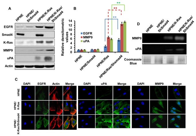
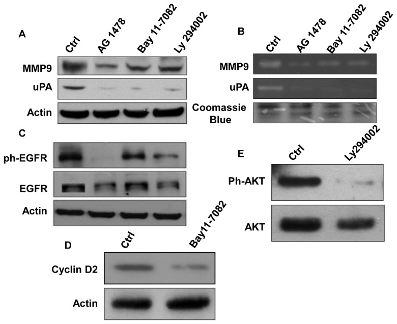
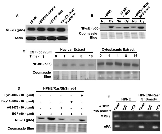
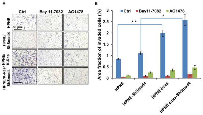
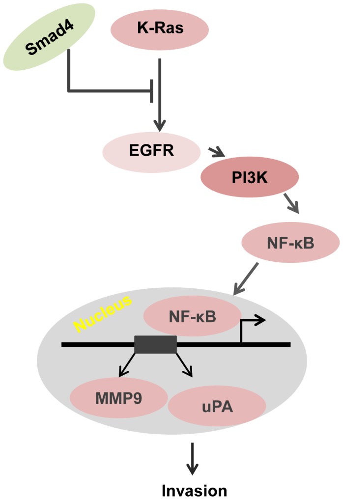
Activating K-Ras mutations and inactivating mutations of Smad4 are two common genetic alterations that occur in the development and progression of pancreatic ductal adenocarcinomas (PDAC). To further study the individual and combinatorial roles of these two mutations in the pathogenesis of PDAC, immortalized human pancreas nestin postive cells (HPNE) were genetically modified by either expressing oncogenic K-Ras (HPNE/K-Ras), by shRNA knock down of Smad4 (HPNE/ShSmad4) or by creating both alterations in the same cell line (HPNE/K-Ras/ShSmad4). We previously found that expression of oncogenic K-Ras caused an increase in expression of EGFR and loss of Smad4 further enhanced the up regulation in expression of EGFR and that this increase in EGFR was sufficient to induce invasion. Here we further investigated the mechanism that links mutational alterations and EGFR expression with invasion. The increase in EGFR signaling was associated with up regulation of MMP9 and uPA protein and activity. Moreover, the increase in EGFR signaling promoted a nuclear translocation and binding of RelA (p65), a subunit of NF-κB, to the promoters of both MMP-9 and uPA. Treatment of HPNE/K-Ras/ShSmad4 cells with an inhibitor of EGFR reduced EGF-mediated NF-κB nuclear translocation and inhibitors of either EGFR or NF-κB reduced the increase in MMP-9 or uPA expression. In conclusion, this study provides the mechanism of how a combination of oncogenic K-Ras and loss of Smad4 causes invasion and provides the basis for new strategies to inhibit metastases.


Conflict of interest statement

Competing Interests: The authors have declared that no competing interests exist.

Figures

Figure 1
Figure 1. Characterization of genetically modified HPNE cell lines.
A. Expression of oncogenic K-Ras and knockdown of Smad4 cooperate to induce EGFR, MMP9 and uPA expressions in HPNE and other K-Ras and Smad4 modified HPNE cells. B. The densitometry values were presented as the measurement of EGFR, MMP9 and uPA expression levels. The values were determined from multiple Western blots (n=3) analyses compared to the density of β-actin that was used as a loading control. The relative expression level of a particular protein was calculated by comparison to the density of the same protein in HPNE cells. Statistical significance value **p< 0.01 was calculated using student’s T-tests. C. Immunostaining followed by images from fluorescent microscopy showed the expression of EGFR, MMP9 and uPA in HPNE and genetically modified HPNE cells. Nuclei were visualized by staining with DAPI (blue). D. Samples (secreted proteins in serum free medium) were collected after 16 hours incubation of the cells in serum free medium. Gelatin and casein zymography analyses were performed to determine MMP9 and uPA activity.
Figure 2
Figure 2. Expression level and activity of MMP9 and uPA was determined after treating HPNE/K-Ras/ShSmad4 cells with different inhibitors.
A. Western blots analysis of MMP9 and uPA after treating cells with AG1478 (10 μM), Bay11-7082 (10 μM) or Ly294002 (10 μM). B. These inhibitors also blocked the secreted levels of MMP9 and uPA as determined by enzymatic activity in gelatin and casein zymography. C. Inhibition of the expression level of ph-EGFR by ph-EGFR, NF-κB and PI3K blockers, AG1478, Bay11-7082 and Ly294002 respectively at above concentrations. D. Western blot analysis was performed to monitor the cyclin D2 expression for the HPNE/K-Ras/ShSmad4 cells treated with Bay11-7082 to confirm the specific effect of Bay11-7082 [42] on NF-κB signaling. E. Western block showing that Ly294002 (10 μM) blocks AKT phosphorylation.
Figure 3
Figure 3. Nuclear translocation of NF-κB sub-unit.
A. Western blot analysis for the NF-κB (p65) expression level in different genetically modified HPNE cells. B. Western blot analysis showing constitutive nuclear expression of p65 in cells expressing oncogenic K-Ras and loss of Smad4. C. Effect of EGF on nuclear translocation of NF-κB (p65). HPNE/K-Ras/ShSmad4 cells are treated with EGF (50 ng/ml) and harvested at indicated time points. Western blot analyses were performed with both nuclear and cytoplasmic extracts. A parallel Coomassie blue stained gel was presented as a loading control. D. Nuclear translocation of p65 is partially blocked by treatment of inhibitors described above in Figure 2. E. NF-B mediated regulation of uPA and MMP9. ChIP assay was performed to show the binding of NF-κB to the promoters of uPA and MMP9. The details for ChIP assay and PCR primers are described in material and methods section.
Figure 4
Figure 4. Loss of Smad4 and expression of oncogenic K-Ras induces invasion in HPNE cells.
A. Images (10X magnification) were taken after 24 hours of seeding the cells in invasion chamber (BD Matrigel Matrix). B. Quantitative analyses were performed using Image J particle analysis program. Five individual images were taken from each chamber and then particle analysis was performed. Fraction of area occupied by the total particles (here indicates the invaded cells) are calculated by this analysis. Bars represent the standard deviation of five different images. Statistical significance of the data is presented as *p<0.05; **p< 0.01 using student’s T-test.
Figure 5
Figure 5. A working model is illustrating how oncogenic K-Ras and loss of Smad4 cooperate to cause an invasive phenotype.
Oncogenic K-Ras signaling induces EGFR expression and K-Ras induced EGFR expression is normally suppressed by Smad4-dependent TGF-β signaling. Loss of Smad4 therefore leads to optimal up-regulation of K-ras induced EGFR expression. The increased expression and signaling by EGFR activates PI3K which induces nuclear translocation of NF-κB that in turn drives the expression of MMP9 and uPA.

Similar articles

Cited by

References

    1. Jemal A, Siegel R, Ward E, Hao Y, Xu J et al. (2008) Cancer statistics, 2008. CA Cancer J Clin 58: 71-96. doi:10.3322/CA.2007.0010. PubMed: 18287387. - DOI - PubMed
    1. Schmalfeldt B, Prechtel D, Härting K, Späthe K, Rutke S et al. (2001) Increased expression of matrix metalloproteinases (MMP)-2, MMP-9, and the urokinase-type plasminogen activator is associated with progression from benign to advanced ovarian cancer. Clin Cancer Res 7: 2396-2404. PubMed: 11489818. - PubMed
    1. Deryugina EI, Quigley JP (2006) Matrix metalloproteinases and tumor metastasis. Cancer Metastasis Rev 25: 9-34. doi:10.1007/s10555-006-7886-9. PubMed: 16680569. - DOI - PubMed
    1. Duffy MJ (2002) Urokinase plasminogen activator and its inhibitor, PAI-1, as prognostic markers in breast cancer: from pilot to level 1 evidence studies. Clin Chem 48: 1194-1197. PubMed: 12142372. - PubMed
    1. Lynch HT, Brand RE, Lynch JF, Fusaro RM, Kern SE (2002) Hereditary factors in pancreatic cancer. J Hepatobiliary Pancreat Surg 9: 12-31. doi:10.1007/s005340200001. PubMed: 12021894. - DOI - PubMed

Publication types

MeSH terms