. 2013 Dec 10;8(12):e83036.
doi: 10.1371/journal.pone.0083036. eCollection 2013.

Plasmablasts as migratory IgG-producing cells in the pathogenesis of neuromyelitis optica

Affiliations

Plasmablasts as migratory IgG-producing cells in the pathogenesis of neuromyelitis optica

Norio Chihara et al. PLoS One. .

Abstract

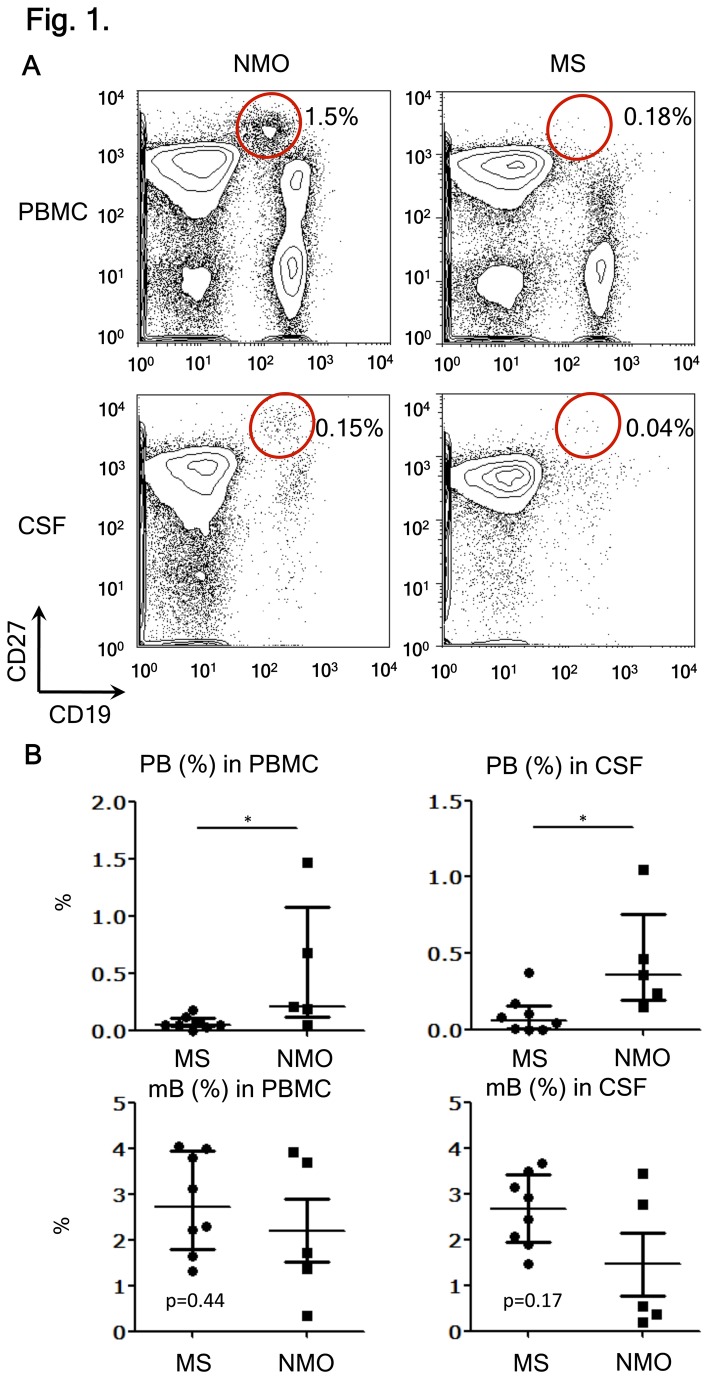
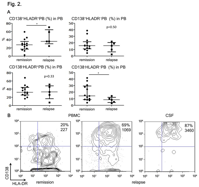
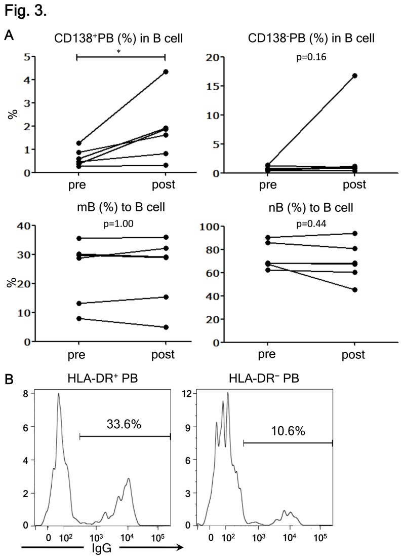
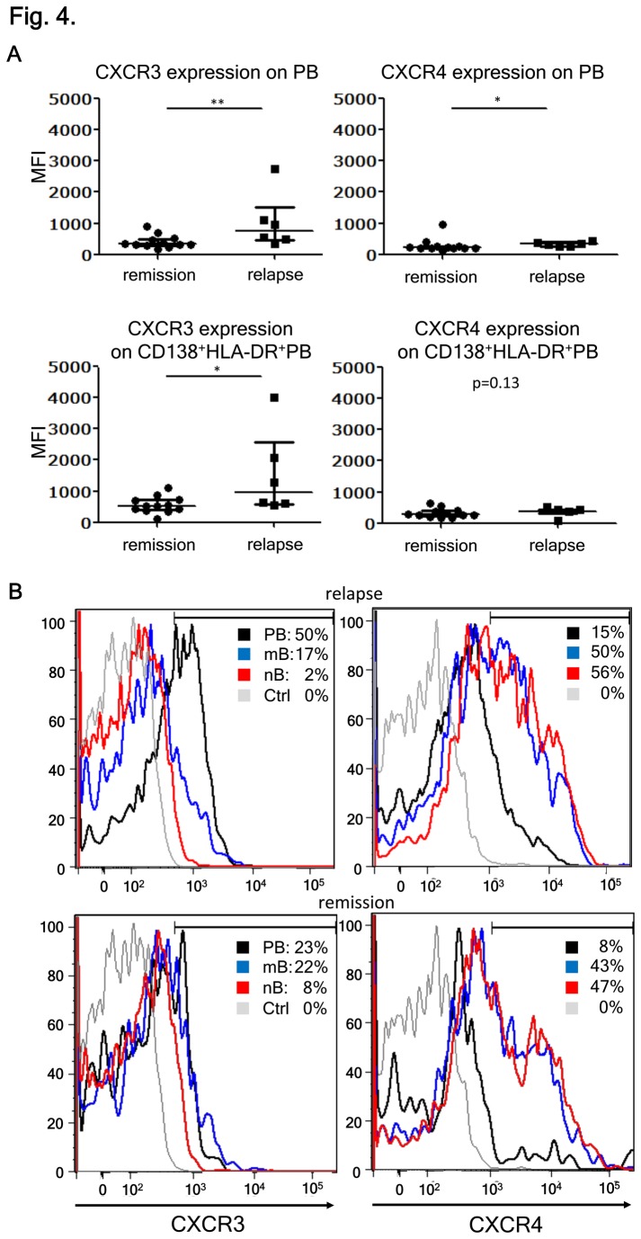
Neuromyelitis optica (NMO) is an inflammatory disease characterized by recurrent attacks of optic neuritis and myelitis. It is generally accepted that autoantibodies against aquaporin 4 water channel protein play a pathogenic role in neuromyelitis optica. We have recently reported that plasmablasts are increased in the peripheral blood of this autoimmune disease, and are capable of producing autoantibodies against aquaporin 4. Here, we demonstrate that CD138(+)HLA-DR(+) plasmablasts, a subset of IgG-producing cells, are increased in the peripheral blood and are enriched among the cerebrospinal fluid (CSF) lymphocytes during the relapse of neuromyelitis optica. Notably, these CD138(+)HLA-DR(+) plasmablasts overexpress CXCR3, whose ligands are present in the cerebrospinal fluid during the relapse of neuromyelitis optica. These results led us to speculate that plasmablasts producing anti-aquaporin 4 autoantibodies might traffic toward the central nervous system (CNS). Furthermore, we performed single-cell sorting of plasmablasts from peripheral blood and CSF samples from NMO and sequenced the complementarity-determining regions (CDRs) of the IgG heavy chain expressed by the sorted plasmablast clones. There were high frequencies of mutations in the CDRs compared with framework regions, indicating that these plasmablast clones would represent a post-germinal center B-cell lineage. Consistent with the preceding results, the plasmablast clones from the peripheral blood shared the same CDR sequences with the clones from the CSF. These results indicate that IgG-producing plasmablasts, which are guided by helper T-cells, may migrate from the peripheral blood preferentially to the CSF. Since migratory plasmablasts could be involved in the inflammatory pathology of NMO, the B-cell subset and their migration might be an attractive therapeutic target.


Conflict of interest statement

Competing Interests: TY serves as a guest editor (reviewer) for this journal. However, this does not alter the authors' adherence to all the PLOS ONE policies on sharing data and materials.

Figures

Figure 1
Figure 1. Selective increase of plasmablasts (PBs) during relapse of NMO.
(A) B-cell subpopulation analysis by flow cytometry. Peripheral blood mononuclear cell (PBMC) and cerebrospinal fluid (CSF) cells were obtained during relapse of neuromyelitis optica (NMO) or multiple sclerosis (MS) and were stained with fluorescence-conjugated anti-CD19, -CD27, -CD38, and -CD180 monoclonal antibodies (mAbs). PB cells (CD19intCD27high) were encircled after observing that they also bear the phenotype of CD38highCD180 (Figure S1). Values represent the percentages of PB cells among all mononuclear cells. (B) The proportion of PB and memory B-cells (mB) in PBMC and CSF from MS and NMO during relapse. The data were obtained from eight patients with MS and five with NMO [*p < 0.05 by Mann–Whitney test; each error bar represents the median ± interquartile range (IQR)].
Figure 2
Figure 2. Kinetics of CD138+HLA-DR+ PB during relapse of NMO.
(A) Analysis of pooled peripheral blood mononuclear cells (PBMC) data from neuromyelitis optica (NMO) in remission and relapse. The plasmablasts (PBs) were subdivided into four subpopulations by considering the expression of CD138 and HLA-DR. The individual data show the percentages of each PB subpopulation among the total PB [*p < 0.05 by Mann–Whitney test; each error bar represents the median ± interquartile range (IQR)]. (B) Enrichment of CD138+HLA-DR+ PBs in the CSF. The PBMC and CSF cells were obtained from NMO during relapse. The values indicate the percentages of CD138+HLA-DR+ PBs among the total PB. The expression level of CD138 was assessed by mean fluorescence intensity (MFI). Representative data of one out of three different cases are shown.
Figure 3
Figure 3. CD138+HLA-DR+ plasmablasts (PB) cells are recently differentiated IgG-producing PB.
(A) The effects of influenza vaccination on the frequencies of B-cell subpopulations were analyzed. The frequency of each B-cell subpopulation derived from the peripheral blood of healthy subjects before (pre) and seven days after vaccination (post) is shown. Each line connects the values obtained from a single subject (*p < 0.05 by Wilcoxon signed rank test). (B) The results of intracellular IgG staining of HLA-DR+ PB (left) and HLA-DR- PBs (right) are shown. The values represent the percentages of IgG-producing cells in each PB subpopulation. Representative data of one out of three individuals are displayed.
Figure 4
Figure 4. CXCR3 expression on plasmablasts (PBs) correlates with the disease state.
(A) CXCR3 and CXCR4 on PB in neuromyelitis optica (NMO) relapse and remission. Here, we compared the mean fluorescence intensity (MFI) of CXCR3 and CXCR4 expression in the peripheral blood PBs during remission and relapse of NMO. MFI of CXCR3 and CXCR4 expressions on CD138+HLA-DR+ PBs were also analyzed [**p < 0.01 and *p < 0.05 by Mann–Whitney test; each error bar represents the median ± interquartile range (IQR]]. (B) B-cell subpopulations derived from peripheral blood mononuclear cells (PBMC) during disease relapse and remission were analyzed by flow cytometry to investigate the expression of CXCR3 and CXCR4. The values represent the percentages of CXCR3+ or CXCR4+ cells within each B-cell subpopulation. Unstained control of PBMC is indicated by Ctrl. Representative data of at least five patients in each disease state are shown.

References

    1. Wingerchuk DM, Hogancamp WF, O'Brien PC, Weinshenker BG (1999) The clinical course of neuromyelitis optica (Devic's syndrome). Neurology 53: 1107-1114. doi:10.1212/WNL.53.5.1107. PubMed: 10496275. - DOI - PubMed
    1. Lennon VA, Wingerchuk DM, Kryzer TJ, Pittock SJ, Lucchinetti CF et al. (2004) A serum autoantibody marker of neuromyelitis optica: distinction from multiple sclerosis. Lancet 364: 2106-2112. doi:10.1016/S0140-6736(04)17551-X. PubMed: 15589308. - DOI - PubMed
    1. Wingerchuk DM, Lennon VA, Lucchinetti CF, Pittock SJ, Weinshenker BG (2007) The spectrum of neuromyelitis optica. Lancet Neurol 6: 805-815. doi:10.1016/S1474-4422(07)70216-8. PubMed: 17706564. - DOI - PubMed
    1. Warabi Y, Matsumoto Y, Hayashi H (2007) Interferon beta-1b exacerbates multiple sclerosis with severe optic nerve and spinal cord demyelination. J Neurol Sci 252: 57-61. doi:10.1016/j.jns.2006.10.008. PubMed: 17125797. - DOI - PubMed
    1. Kleiter I, Hellwig K, Berthele A, Kümpfel T, Linker RA et al. (2012) Neuromyelitis Optica Study Group. Failure of natalizumab to prevent relapses in neuromyelitis optica. Arch Neurol 69: 239-245. doi:10.1001/archneurol.2011.216. PubMed: 22332191. - DOI - PubMed

Publication types

MeSH terms