. 2013 Dec 16;6(1):182.
doi: 10.1186/1754-6834-6-182.

A synthetic biology approach for evaluating the functional contribution of designer cellulosome components to deconstruction of cellulosic substrates

Affiliations

A synthetic biology approach for evaluating the functional contribution of designer cellulosome components to deconstruction of cellulosic substrates

Yael Vazana et al. Biotechnol Biofuels. .

Abstract

Background: Select cellulolytic bacteria produce multi-enzymatic cellulosome complexes that bind to the plant cell wall and catalyze its efficient degradation. The multi-modular interconnecting cellulosomal subunits comprise dockerin-containing enzymes that bind cohesively to cohesin-containing scaffoldins. The organization of the modules into functional polypeptides is achieved by intermodular linkers of different lengths and composition, which provide flexibility to the complex and determine its overall architecture.

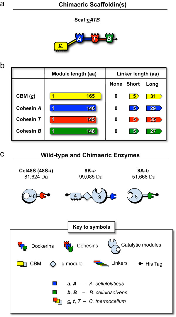
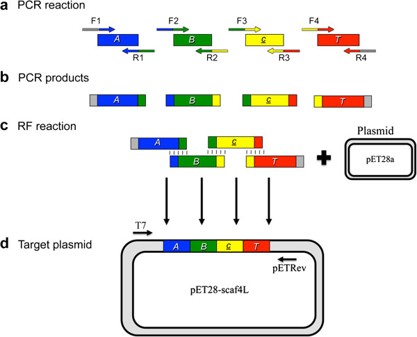
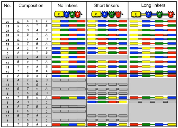
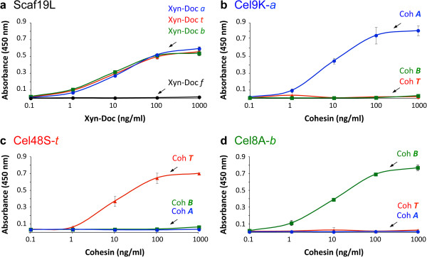
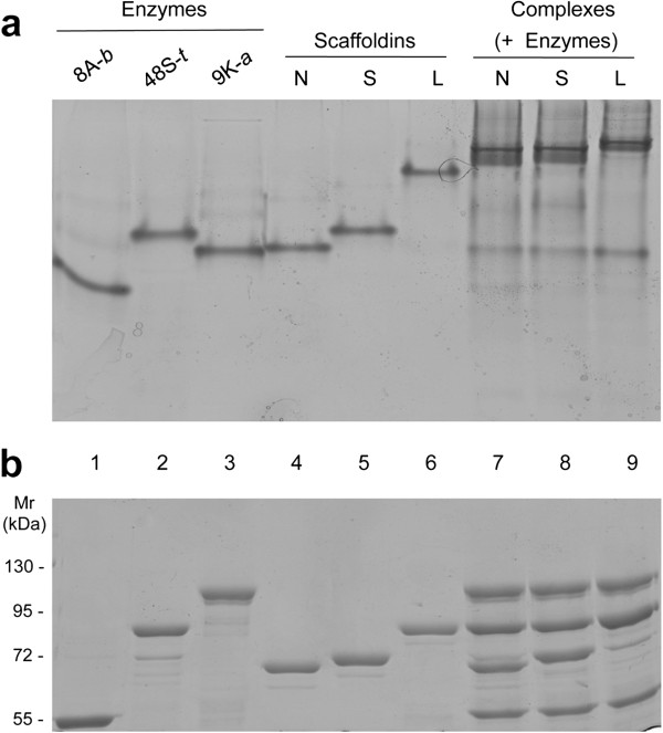
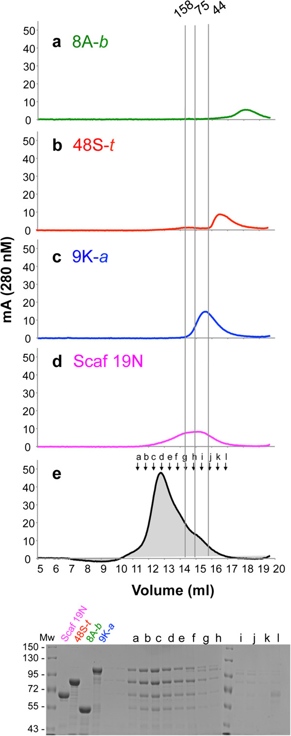
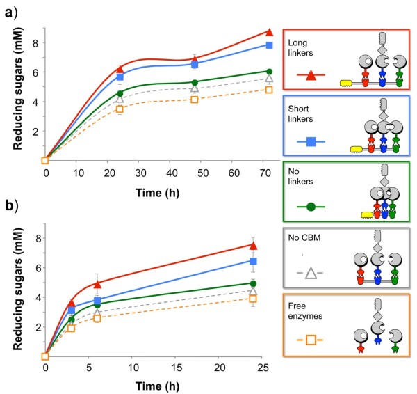
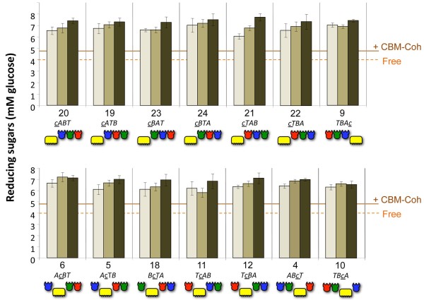
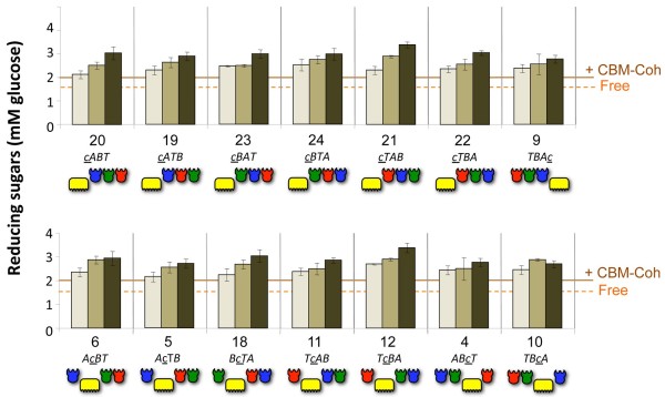
Results: Using a synthetic biology approach, we systematically investigated the spatial organization of the scaffoldin subunit and its effect on cellulose hydrolysis by designing a combinatorial library of recombinant trivalent designer scaffoldins, which contain a carbohydrate-binding module (CBM) and 3 divergent cohesin modules. The positions of the individual modules were shuffled into 24 different arrangements of chimaeric scaffoldins. This basic set was further extended into three sub-sets for each arrangement with intermodular linkers ranging from zero (no linkers), 5 (short linkers) and native linkers of 27-35 amino acids (long linkers). Of the 72 possible scaffoldins, 56 were successfully cloned and 45 of them expressed, representing 14 full sets of chimaeric scaffoldins. The resultant 42-component scaffoldin library was used to assemble designer cellulosomes, comprising three model C. thermocellum cellulases. Activities were examined using Avicel as a pure microcrystalline cellulose substrate and pretreated cellulose-enriched wheat straw as a model substrate derived from a native source. All scaffoldin combinations yielded active trivalent designer cellulosome assemblies on both substrates that exceeded the levels of the free enzyme systems. A preferred modular arrangement for the trivalent designer scaffoldin was not observed for the three enzymes used in this study, indicating that they could be integrated at any position in the designer cellulosome without significant effect on cellulose-degrading activity. Designer cellulosomes assembled with the long-linker scaffoldins achieved higher levels of activity, compared to those assembled with short-and no-linker scaffoldins.

Conclusions: The results demonstrate the robustness of the cellulosome system. Long intermodular scaffoldin linkers are preferable, thus leading to enhanced degradation of cellulosic substrates, presumably due to the increased flexibility and spatial positioning of the attached enzymes in the complex. These findings provide a general basis for improved designer cellulosome systems as a platform for bioethanol production.


Figures

Figure 1
Figure 1
Schematic representation of the recombinant proteins used in this study. The modular notation, structure and molecular mass of each protein are indicated. Red, yellow and light blue indicate C. thermocellum-derived cohesin/dockerin, carbohydrate binding module (CBM) and enzyme-related components, respectively. Dark blue indicates A. cellulolyticus-derived cohesin/dockerin modules, and green indicates B. cellulosolvens-derived modules. (a) The basic chimaeric scaffoldin containing three divergent cohesins: the third cohesin of scaffoldin ScaC from A. cellulolyticus (A), the third cohesin of ScaB from B. cellulosolvens (B), the second cohesin of the CipA scaffoldin from C. thermocellum (T) plus a CBM3a module of the same scaffoldin (c). See Additional file 1: Table S1 for the molecular weights of the respective chimaeric scaffoldins. (b) The length of each module and its C-flanking linkers in amino acid residues. (c) Recombinant cellulases used in this study. In the modular notation of the enzymes, the number indicates the GH family of the catalytic domain. S, K and A indicate the original name of the enzyme (Cel48S, Cel9K and Cel8A, respectively). The chimaeric Cel9K includes a CBM4 and Ig domain on the N-terminal portion of the enzyme. Lowercase t, a and b indicate the source of the dockerin module (C. thermocellum, A. cellulolyticus and B. cellulosolvens respectively).
Figure 2
Figure 2
Multi-component assembly of a chimaeric scaffoldin - ‘Scaf4L’. (a) Schematic representation of the four components that were amplified in the first PCR reaction: A. cellulolyticus cohesin (A) and C-terminal linker segment, B. cellulosolvens cohesin (B) and linker, carbohydrate binding module (CBM) (c) and linker and C. thermocellum cohesin (T) but without the C-terminal linker. (b) Resultant PCR products. (c) The PCR products were then used as mega-primers for the restriction-free reaction with the pET28a plasmid. The sequences that form base-pairing due to flanking regions designed in the primers of each module are indicated by the coloring of the PCR products and by connecting lines. (d) The resulting linear plasmid pET28a-scaf4L. Ligation occurs spontaneously in E. coli.
Figure 3
Figure 3
Schematic representation of the scaffoldins in the final scaffoldin library. Twenty-four different arrangements of the cohesin (A, B and T) and carbohydrate binding module (CBM) (c) modules are shown in three sub-libraries: no-linker, short-linker and long-linker versions of the given chimaeric scaffoldins. The left column indicates the number of each scaffoldin set according to its composition (position of CBM and divergent cohesins). The 45 successfully cloned and expressed scaffoldins included in the final library are shown as colored pictograms. An additional 11 scaffoldins were cloned but not expressed (shown as gray pictograms); 16 additional scaffoldins were not expressed (gray background); 14 full sets, representing 42 cloned and expressed scaffoldins, were finally achieved for further study.
Figure 4
Figure 4
Specificity of the cohesin-borne scaffoldin for its matching dockerins (a) and of the dockerin-bearing enzymes for their target cohesins (b-d). The interaction between scaffoldin 19L (Scaf19L) and its matching dockerins was examined (a) using a standardized matching fusion-protein system [73]. Scaf19L was coated onto the wells of a microtiter plate and was subjected to interaction with Xyn-Doc fusion proteins, Xyn-Doc t (red), Xyn-Doc a (blue), Xyn-Doc b (green), and a non-matching control Xyn-Doc f, the divergent dockerin of which was derived from a cellulosomal component of the rumen bacterium, Ruminococcus flavefaciens (black). Anti-xylanase antibodies were used, together with a second antibody-conjugated enzyme system to promote a chromogenic reaction. Similarly, the interaction between the enzymes via their dockerin module to matching and non-matching cohesins was examined. In this case, 9K-a (b), 48S-t (c) and 8A-b (d), which bear an A. cellulolyticus, C. thermocellum and B. cellulosolvens dockerin, respectively, were coated onto the wells of microtiter plates and subjected to interaction with the designated carbohydrate binding module (CBM)-fused cohesin modules, Coh T (red), Coh A (blue) and Coh B (green).
Figure 5
Figure 5
Electrophoretic mobility of components and assembled complexes on non-denaturing PAGE (a) and SDS-PAGE (b). Equimolar concentrations of the enzymes 48S-t, 9K-a and 8A-b (lanes 1 to 3 respectively) and the matching scaffoldins without intermodular linkers, with short and long intermodular linkers (Scaf19N, Scaf19S and Scaf19L designated as: N, S and L, lanes 4 to 6, respectively) were interacted to form the respective designer cellulosome complexes (lanes 6 to 9). Mr, molecular mass marker.
Figure 6
Figure 6
Superdex 200 gel filtration fast protein liquid chromatography (FPLC) elution profile of a designer-cellulosome complex composed of three chimaeric C. thermocellum enzymes, 9K-a, 48S-t and 8A-b, assembled on a trivalent chimaeric scaffoldin (Scaf19N) without intermodular linkers. The elution profile of each of the single components was used as a marker. The curves are labeled as follows: (a) 8A-b: green, 51.6 kDa, (b) 48S-t: red, 81.6 kDa|, (c) 9K-a: blue, 101.4 kDa, (d) scaffoldin 19N: magenta, 66 kDa, and (e) designer cellulosome complex: black with gray filling. The gel on the bottom shows the SDS-PAGE analysis of the designated elution fractions of the designer cellulosome complexes.
Figure 7
Figure 7
Kinetics of Avicel (a) and pretreated cellulose-enriched wheat straw (b) hydrolysis by designer-cellulosome complexes and free enzymes. The graphs show degradation by scaffoldin-set 21 with the following modular organization cTAB: long-linker scaffoldin-based designer-cellulosome (red), the short linker-based designer cellulosomes (blue), and the designer cellulosomes based on the scaffoldin without intermodular linkers (green). Controls include degradation by a designer cellulosome containing a scaffoldin that lacks a carbohydrate binding module (CBM) (gray) and degradation by the free enzymes (orange). Enzymatic activity was defined by release of reducing sugars (mM) as determined by a glucose standard curve. All reactions were carried out in triplicate. Standard deviations from three separate experiments are indicated.
Figure 8
Figure 8
Comparative hydrolysis of Avicel by the 14 sets of designer cellulosomes. The modular composition of each set and the scaffoldin number is denoted on the x-axis. Upper panel: the carbohydrate binding module (CBM) of the designer scaffoldin is at one of the terminal positions (N or C terminus). Lower panel: the CBM module of the designer scaffoldin is in an internal position. Each designer-cellulosome set is assembled either without intermodular linker scaffoldin (light brown), with short intermodular linker scaffoldin (medium brown), and with long intermodular linker scaffoldin (dark brown). Controls: Free: corresponds to the combined activity of 48S-t, 9K-a and 8A-b. CBM-Coh represents a cellulose-targeting control, corresponding to the activity of the three dockerin-bearing enzymes, each attached separately to its matching cohesin module fused to a CBM. Reactions were carried out for 72 h. Enzymatic activity was defined by mM reducing sugars as determined by a glucose standard curve. All reactions were carried out in triplicate and repeated three times. Standard deviations of at least three experiments are indicated.
Figure 9
Figure 9
Comparative hydrolysis of pretreated cellulose-enriched wheat straw by the 14 sets of designer cellulosomes. Reactions were carried out for 3 h on pretreated cellulose-enriched wheat straw. All other details are provided in the legend to Figure 8.

Similar articles

Cited by

References

    1. Lamed R, Setter E, Bayer EA. Characterization of a cellulose-binding, cellulase-containing complex in Clostridium thermocellum. J Bacteriol. 1983;6:828–836. - PMC - PubMed
    1. Lamed R, Setter E, Kenig R, Bayer EA. The cellulosome - a discrete cell surface organelle of Clostridium thermocellum which exhibits separate antigenic, cellulose-binding and various cellulolytic activities. Biotechnol Bioeng Symp. 1983;6:163–181.
    1. Dassa B, Borovok I, Lamed R, Henrissat B, Coutinho P, Hemme CL, Huang Y, Zhou J, Bayer EA. Genome-wide analysis of Acetivibrio cellulolyticus provides a blueprint of an elaborate cellulosome system. BMC Genomics. 2012;6:210. doi: 10.1186/1471-2164-13-210. - DOI - PMC - PubMed
    1. Belaich JP, Tardif C, Belaich A, Gaudin C. The cellulolytic system of Clostridium cellulolyticum. J Biotechnol. 1997;6:3–14. doi: 10.1016/S0168-1656(97)00085-0. - DOI - PubMed
    1. Xu Q, Bayer EA, Goldman M, Kenig R, Shoham Y, Lamed R. Architecture of the Bacteroides cellulosolvens cellulosome: description of a cell surface-anchoring scaffoldin and a family 48 cellulase. J Bacteriol. 2004;6:968–977. doi: 10.1128/JB.186.4.968-977.2004. - DOI - PMC - PubMed