. 2014 Apr 15;31(8):773-81.
doi: 10.1089/neu.2013.3181. Epub 2014 Feb 7.

Evidence for the therapeutic efficacy of either mild hypothermia or oxygen radical scavengers after repetitive mild traumatic brain injury

Affiliations

Evidence for the therapeutic efficacy of either mild hypothermia or oxygen radical scavengers after repetitive mild traumatic brain injury

Takashi Miyauchi et al. J Neurotrauma. .

Abstract

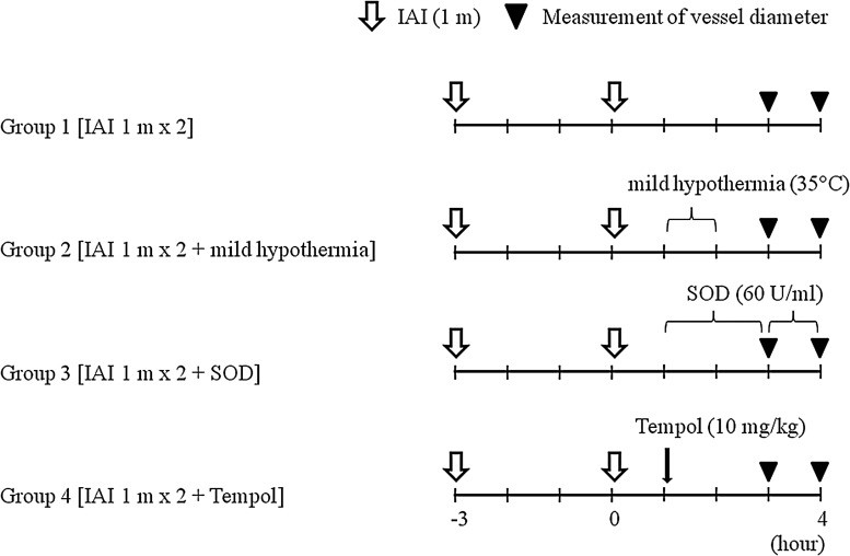
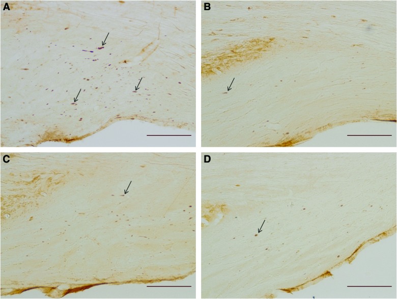
Repetitive brain injury, particularly that occurring with sporting-related injuries, has recently garnered increased attention in both the clinical and public settings. In the laboratory, we have demonstrated the adverse axonal and vascular consequences of repetitive brain injury and have demonstrated that moderate hypothermia and/or FK506 exerted protective effects after repetitive mild traumatic brain injury (mTBI) when administered within a specific time frame, suggesting a range of therapeutic modalities to prevent a dramatic exacerbation. In this communication, we revisit the utility of targeted therapeutic intervention to seek the minimal level of hypothermia needed to achieve protection while probing the role of oxygen radicals and their therapeutic targeting. Male Sprague-Dawley rats were subjected to repetitive mTBI by impact acceleration injury. Mild hypothermia (35 °C, group 2), superoxide dismutase (group 3), and Tempol (group 4) were employed as therapeutic interventions administered 1 h after the repetitive mTBI. To assess vascular function, cerebral vascular reactivity to acetylcholine was evaluated 3 and 4 h after the repetitive mTBI, whereas to detect the burden of axonal damage, amyloid precursor protein (APP) density in the medullospinal junction was measured. Whereas complete impairment of vascular reactivity was observed in group 1 (without intervention), significant preservation of vascular reactivity was found in the other groups. Similarly, whereas remarkable increase in the APP-positive axon was observed in group 1, there were no significant increases in the other groups. Collectively, these findings indicate that even mild hypothermia or the blunting free radical damage, even when performed in a delayed period, is protective in repetitive mTBI.


Figures

<b>FIG. 1.</b>
FIG. 1.
This chart shows the time course for each experimental group. All animals were exposed to twice repetitive impact acceleration insults at 3-h intervals. In group 1, no intervention was administered to animals. In group 2, mild hypothermia (35°C) was initiated 1 h after repetitive mTBI and rectal temperature was maintained at 35°C for 1 h, followed by gradual rewarming. In group 3, superoxide dismutase (SOD; 60 U/mL) was administered into artificial cerebrospinal fluid (aCSF)-containing space underlying the cranial window 1 h after repetitive mTBI. Cerebral vascular was exposed to SOD solution for 3 h in total. In group 4, Tempol (10 mg/kg) was administered by the catheter placed in the femoral vein 1 h after repetitive mTBI. Vascular reactivity to acetylcholine was assessed at 3 and 4 h after repetitive mTBI. mTBI, mild traumatic brain injury; IAI, impact acceleration insult.
<b>FIG. 2.</b>
FIG. 2.
This figure illustrates changes of mean rectal temperatures throughout the duration of the study. A parallel temperature response was observed with the use of a temporalis muscle probe, with the caveat that this temperature slightly trailed the rectal temperature by approximately 0.5°C. Rectal temperature in group 2 was decreased quickly and maintained at 35°C for 1 h. Rewarming was performed gradually and temperature achieved the targeted range 1 h later. Arrow indicates the time of each insult. Data points represent 10-min intervals. Timeline represents the time course after the first insult. Values are expressed as mean±standard error of the mean.
<b>FIG. 3.</b>
FIG. 3.
These bar graphs show cerebral vascular reactivity to 10−7 and 10−5 M ACh 3 and 4 h after repetitive mTBI. Bars express percent changes from baseline cerebral vascular diameter after exposure to ACh in each measurement time point. In group 1, cerebral vascular responses were significantly suppressed at all measurement points (left panel). In group 2, cerebral vascular responses were significantly higher than in group 1 in all measurement points (center panel). In groups 3 and 4, cerebral vascular responses were also significantly higher than group 1 in all measurement points (right panel). Vascular responses to 10−5 M ACh at 3 and 4 h after the repetitive mTBI in group 3 were significantly higher than group 4. All values are expressed as mean±standard error of the mean. Statistical difference was analyzed by Kruskal-Wallis' test followed by Bonferroni's test for multiple comparisons. *Significant difference at p<0.001 versus group 1; #significant difference at p<0.05. ACh, acetylcholine.
<b>FIG. 4.</b>
FIG. 4.
Representative micrographs of medullospinal junction in each group. Brain stem was obtained 4 h after repetitive mTBI. Bulb-shaped black dots (arrows) are amyloid precursor protein (APP)-positive axons, suggesting significant axonal damage and disconnection. Significantly increased APP-positive axons can be observed in group 1 (A), whereas decreased numbers of APP-positive profiles are shown in the remaining groups (B, C, and D). Original magnification, 100×. Bar, 200 μm.
<b>FIG. 5.</b>
FIG. 5.
This bar graph shows mean density of APP-immunoreactive damaged axon in the medullospinal tract at 4 h after repetitive mTBI. Significantly increased APP density was observed in group 1. Whereas APP density in groups 2, 3, and 4 were significantly lower than that in group 1, there were no significant differences between therapeutic intervention groups (groups 2, 3, and 4). All values represent the mean±standard error of the mean. Statistical differences were analyzed by the Kruskal-Wallis' test, followed by Bonferroni's test for multiple comparisons. *Significant difference at p<0.001.

References

    1. Allen G.V., Gerami D., and Esser M.J. (2000). Conditioning effects of repetitive mild neurotrauma on motor function in an animal model of focal brain injury. Neuroscience 99, 93–105 - PubMed
    1. Laurer H., Bareyre F.M., Lee V.M.Y.C., Trojanowski J.Q., Longhi L., Hoover R., Saatman K.E., Raghupathi R., Hoshino S., Grady M.S., and McIntosh T.K. (2001). Mild head injury increasing the brain's vulnerability to a second concussive impact. J. Neurosurg. 95, 859–870 - PubMed
    1. Collins M.W., Lovell M.R., Iverson G.L., Cantu R.C., Maroon J.C., and Field M. (2002). Cumulative effects of concussion in high school athletes. Neurosurgery 51, 1175–1181 - PubMed
    1. Raghpathi R., Mehr M.F., Helfaer M.A., and Margulies S.S. (2004). Traumatic axonal injury is exacerbated following repetitive closed head injury in the neonatal pig. J. Neurotrauma 21, 307–316 - PubMed
    1. Moser R.S., Schatz P., and Jordan B.D. (2005). Prolonged effects of concussion in high school athletes. Neurosurgery 57, 300–306 - PubMed

Publication types