

Human papillomavirus proteins are found in peripheral blood and semen Cd20+ and Cd56+ cells during HPV-16 semen infection
- PMID: 24341689
- PMCID: PMC3878630
- DOI: 10.1186/1471-2334-13-593
Human papillomavirus proteins are found in peripheral blood and semen Cd20+ and Cd56+ cells during HPV-16 semen infection
Abstract
Background: Human papillomavirus (HPV) currently represents an important risk factor for cancer development and infertility in humans. Whilst binding of HPV to spermatozoa has been associated with male infertility, an investigation about the presence of HPV-DNA in non-spermatozoal semen cells is lacking. Previous findings documented the presence of HPV in peripheral blood leukocytes. The aim of this study was to investigate the expression of HPV markers in semen and blood leukocytes during HPV-16 infection.
Methods: A total of 32 subjects, 16 patients affected by HPV-16 semen infection and 16 controls, were evaluated in our andrological centre and enrolled in the study. Semen non-spermatozoal cells from all subjects were isolated and evaluated for the expression of HPV-16 markers (DNA and L1, E6 proteins) and further characterized for their molecular phenotype. Analogue determination was performed on peripheral blood mononuclear cells.
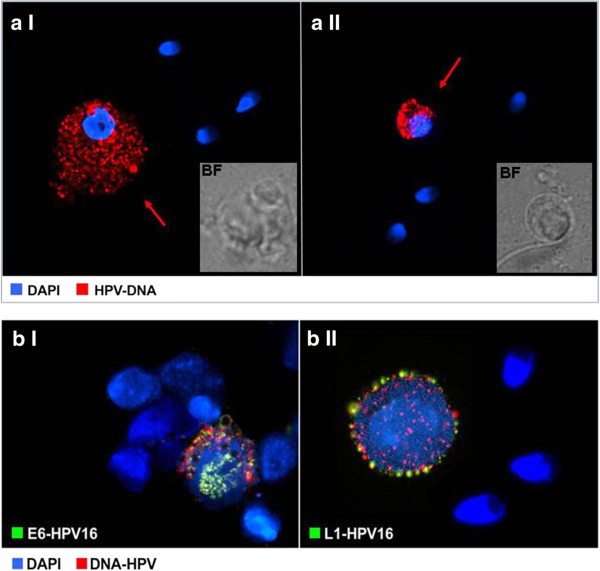
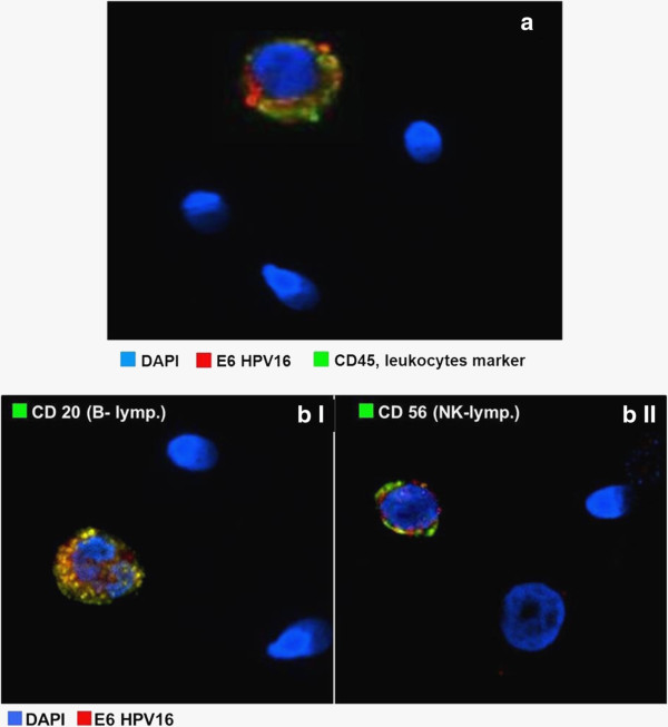
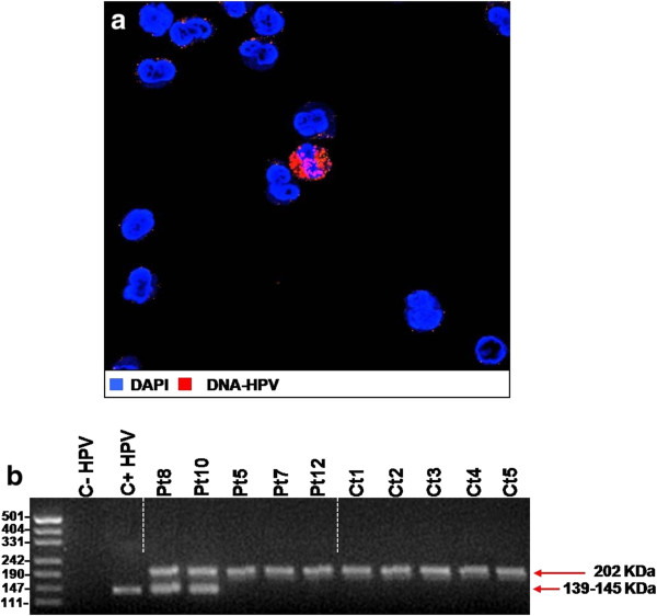
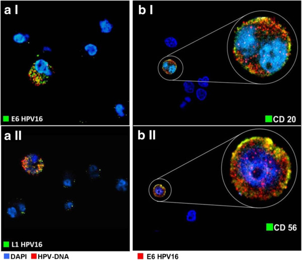
Results: The presence of HPV-DNA by FISH analysis in a round cell population from semen, confirmed to be CD45+ leukocytes, was observed. These HPV-DNA containing-cells also displayed HPV-16-E6 and HPV-16-L1 viral proteins and, upon further investigation, were found to be CD20+ and CD56+, likely phenotypes of B cells and natural killer cells (NK) respectively. In 25% of the patient group, a very small population of peripheral blood mononuclear cells was found to be positive for HPV-DNA via FISH. These cells displayed the CD20+ and CD56+ phenotype alike. None of the control subjects displayed HPV-DNA in either semen or peripheral blood.
Conclusion: Considering the role of CD20+ and CD56+ cell populations in the antiviral immune response, the detection of HPV markers on leukocytes may reflect the presence of virus particles within the endosomal compartment. However, the presence of HPV markers in circulating mononuclear cells raise concerns about the risk of developing cancers to distal organs.
Figures
References
-
- Foresta C, Pizzol D, Moretti A, Barzon L, Palù G, Garolla A. Clinical and prognostic significance of human papillomavirus DNA in the sperm or exfoliated cells of infertile patients and subjects with risk factors. Fertil Steril. 2010;13:1723–1727. doi: 10.1016/j.fertnstert.2009.11.012. - DOI - PubMed
MeSH terms
Substances
LinkOut - more resources
Full Text Sources
Other Literature Sources
Research Materials
Miscellaneous
