. 2014 Jan 14;53(1):135-42.
doi: 10.1021/bi401492k. Epub 2013 Dec 30.

Photoaffinity labeling the propofol binding site in GLIC

Affiliations

Photoaffinity labeling the propofol binding site in GLIC

David C Chiara et al. Biochemistry. .

Abstract

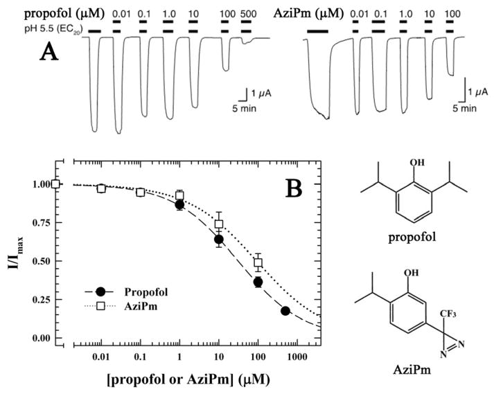
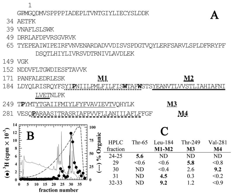
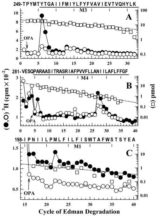
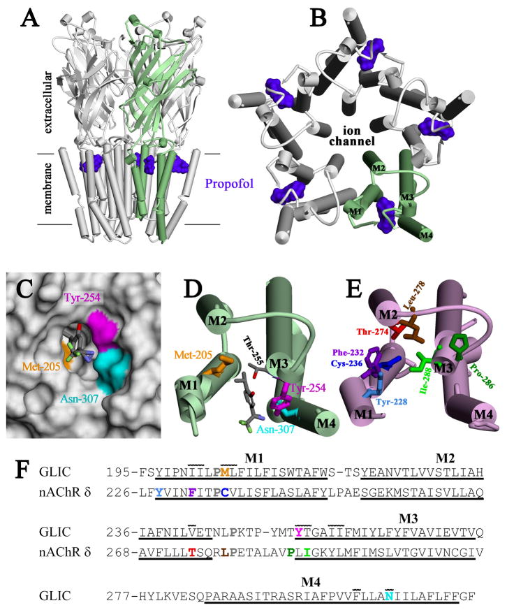
Propofol, an intravenous general anesthetic, produces many of its anesthetic effects in vivo by potentiating the responses of GABA type A receptors (GABAAR), members of the superfamily of pentameric ligand-gated ion channels (pLGICs) that contain anion-selective channels. Propofol also inhibits pLGICs containing cation-selective channels, including nicotinic acetylcholine receptors and GLIC, a prokaryotic proton-gated homologue from Gloeobacter violaceus . In the structure of GLIC cocrystallized with propofol at pH 4 (presumed open/desensitized states), propofol was localized to an intrasubunit pocket at the extracellular end of the transmembrane domain within the bundle of transmembrane α-helices (Nury, H, et al. (2011) Nature 469, 428-431). To identify propofol binding sites in GLIC in solution, we used a recently developed photoreactive propofol analogue (2-isopropyl-5-[3-(trifluoromethyl)-3H-diazirin-3-yl]phenol or AziPm) that acts as an anesthetic in vivo and potentiates GABAAR in vitro. For GLIC expressed in Xenopus oocytes, propofol and AziPm inhibited current responses at pH 5.5 (EC20) with IC50 values of 20 and 50 μM, respectively. When [(3)H]AziPm (7 μM) was used to photolabel detergent-solubilized, affinity-purified GLIC at pH 4.4, protein microsequencing identified propofol-inhibitable photolabeling of three residues in the GLIC transmembrane domain: Met-205, Tyr-254, and Asn-307 in the M1, M3, and M4 transmembrane helices, respectively. Thus, for GLIC in solution, propofol and AziPm bind competitively to a site in proximity to these residues, which, in the GLIC crystal structure, are in contact with the propofol bound in the intrasubunit pocket.


Conflict of interest statement

CONFLICT OF INTEREST DISCLOSURE: The authors declare no competing financial interest.

Figures

Figure 1
Figure 1. Propofol and AziPm both inhibit H+ activated GLIC currents
A) Representative current traces of oocyte-expressed GLIC exposed to pH 5.5 corresponding to the EC20 in the presence of various concentrations of propofol or AziPm as indicated with black bars over the traces. Limited solubility of AziPm prevented analysis at higher concentrations. B) Inhibition curves for propofol and AziPm at the pH corresponding to the EC20 (pH 5.5). Response is expressed as the fraction of current induced in the presence of the indicated concentrations of propofol (●) or AziPm (□) relative to that in their absence. The data (n=7) were fit to Hill equations with IC50s of 21.4 ± 1.2 and 51.4 ± 5.3 μM, respectively, and had Hill coefficients of −0.67 ± 0.05 and −0.63 ± 0.11, respectively. Error bars represent SEM. Also included are the chemical structures of propofol and AziPm.
Figure 2
Figure 2. Reversed-phase HPLC fractionation of EndoLys-C digests of GLIC: [3H]AziPm primarily photoincorporate into the hydrophobic, transmembrane domain of GLIC
A) Shown are the sequences of the GLIC fragments produced by enzymatic cleavage with EndoLys-C (specific for Lys). The residue numbering is that used in the crystal structure (PDB:3P50 (10)) and does not include the 3 additional N-terminal residues (GPM) identified by sequence analysis of intact, expressed GLIC. The transmembrane helices M1–M4 are underlined. The Pro residues used to chemically isolate M1, M3, & M4 during sequencing are bolded as are the Trp residues subjected to chemical cleavage to sequence M2. B) Reversed-phase HPLC fractionation of an EndoLys-C digest of [3H]AziPm labeled GLIC. 82% of the recovered 3H eluted in hydrophobic fractions (>75 % organic, fractions 28–38). In gray is the absorbance profile at 215 nm. C) Selected fractions containing 3H were sequenced, with the peptides detected quantitated in picomoles. In fractions 29–33, the only GLIC fragments detected were those containing the transmembrane helices. ND, not detected.
Figure 3
Figure 3. Propofol-inhibitable [3H]AziPm labeling identified in M3, M4, and M1
3H (●,○) and PTH-amino acids (□) released when equal aliquots of EndoLys-C digests of GLIC photolabeled with [3H]AziPm in the absence (●) or presence (○,□) of 300 μM propofol were sequenced with OPA treatment in cycles 2 (A), 5 (B), or 16 (C). A) After OPA treatment in cycle 2, the primary sequence began at Thr-249 before M3 (I0 = 20 pmol, both conditions). The GLIC amino-terminal fragment, the only other peptide from an EndoLys-C digest of GLIC with a proline in cycle 2, was present (11 pmol, both conditions). The peak of 3H release in cycle 6 indicated of photolabeling of Tyr-254 at an efficiency of 115 cpm/pmol, which 300 μM propofol inhibited by 83%. The small peak of 3H release in cycle 13 indicated propofol-insensitive photoabeling of Met-261 at ~20 cpm/pmol. B) After OPA treatment in cycle 5, the primary sequence began at Val-281 before M4 (-PPF, I0 = 15 pmol; +PPF (□), I0 = 21 pmol). The only other peptide present was lag from the fragment beginning Thr-65 (13 pmol), which contains Pro-68 in cycle 4 that would also be present in cycle 5 as a consequence of the ~90% repetitive yield of Edman degradation. The peak of 3H release in cycle 27 indicated photolabeling of Asn-307 in M4 at 300 cpm/pmol which 300 μM propofol inhibited by 70%. Since treatment with OPA blocks ~90 % of the free amino termini of peptides not containing a Pro, the peak of 3H release in cycle 6 after the OPA treatment is consistent with photolabeling of Tyr-254 if the M3 fragment beginning Thr-249 is present at ~7% the level seen in Panel A. C) After OPA treatment in cycle 16, the primary sequence detected originally began at Leu-184 (-PPF, I0 = 9 pmol; +PPF (□) I0 = 5 pmol). The peak of 3H release in cycle 22 indicated photolabeling of Met-205 at ~40 cpm/pmol, which propofol inhibited by ~40 %.
Figure 4
Figure 4. Location of [3H]AziPm-photolabeled residues in the propofol binding site in GLIC and in the equivalent binding site in the Torpedo nAChR δ subunit
Shown in panels A–D are views of the crystallographic model of GLIC co-crystalized with propofol (PDB:3P50) and in panel E a view of the nAChR δ subunit transmembrane domain (TMD) from an nAChR homology model derived from the GLIC structure (see Methods and Supporting Information Supplemental Figures S1 and S2). α-Helices are shown as cylinders and β-sheets as ribbons. A) A view of GLIC from the side and B) a view of the GLIC TMD from the base of the extracellular domain, with one subunit colored green. The positions of propofol within the structure are shown as Connolly surfaces (purple). C) Connolly surface representation of the propofol binding pocket, viewed from the lipid, with the surfaces contributed by the photolabeled residues color-coded: Tyr-254 (magenta), Asn-307 (cyan), and Met-205 (gold). The lowest energy docking solution for AziPm is included in stick format, color-coded by atom type: gray, carbon; red, oxygen; blue, nitrogen; light green, fluorine. D) A view of a single subunit’s TMD from the same perspective as in B, illustrating the position of the docked AziPm solution and the [3H]AziPm-photolabeled residues in stick format, color-coded as in C. The propofol/AziPm binding pocket resides between the M1, M3 and M4 helices. E) A view of the nAChR δ subunit TMD, oriented similar to GLIC in D. Residues in the δ subunit helix bundle pocket photolabeled by various photoreactive nAChR inhibitors are shown in stick format: [3H]AziPm Phe-232, Cys-236, & Thr-274 (20); [14C]halothane δTyr-228 (33); [125I]TID Ile-288, Phe-232, Cys-236, Thr-274, & Leu-278 (27,34,35); [3H]benzophenone Pro-286, Ile-288, & Phe-232 (28); [3H]azietomidate Cys-236 (41); and [3H]TFD-etomidate Phe-232 & Cys-236 (29). In contrast to the propofol/AziPm site in GLIC, the drug binding pocket in the nAChR δ subunit resides between the M1, M2, & M3 helices. F) The subunit primary structure alignment in the M1–M3 region used to make the nAChR homology model from the GLIC structure. The conserved prolines in all pLGICs are shown in gray. Also included is the M4 sequence from GLIC. The extent of the transmembrane helices is denoted by underlining. Photolabeled residues in GLIC and the Torpedo nAChR δ subunit are color-coded as in D & E. Residues in GLIC in contact with propofol in the crystal structure (10) have a line over them, illustrating the similarities between the intrasubunit pockets of GLIC and Torpedo nAChR δ subunit.

Similar articles

Cited by

References

    1. Krasowski MD, Jenkins A, Flood P, Kung AY, Hopfinger AJ, Harrison NL. General anesthetic potencies of a series of propofol analogs correlate with potency for potentiation of gamma-aminobutyric acid (GABA) current at the GABA(A) receptor but not with lipid solubility. J Pharmacol Exp Ther. 2001;297:338–351. - PubMed
    1. Rudolph U, Antkowiak B. Molecular and neuronal substrates for general anaesthetics. Nat Rev Neurosci. 2004;5:709–720. - PubMed
    1. Franks NP. General anaesthesia: from molecular targets to neuronal pathways of sleep and arousal. Nat Rev Neurosci. 2008;9:370–386. - PubMed
    1. Dilger JP, Vidal AM, Mody HI, Liu Y. Evidence for direct actions of general anesthetics on an ion channel protein - A new look at a unified mechanism of action. Anesthesiology. 1994;81:431–442. - PubMed
    1. Violet JM, Downie DL, Nakisa RC, Lieb WR, Franks NP. Differential sensitivities of mammalian neuronal and muscle nicotinic acetylcholine receptors to general anesthetics. Anesthesiology. 1997;86:866–874. - PubMed

Publication types

MeSH terms

LinkOut - more resources