Review
. 2014 Aug;1843(8):1475-88.
doi: 10.1016/j.bbamcr.2013.12.007. Epub 2013 Dec 14.

Lipids and topological rules governing membrane protein assembly

Affiliations
Review

Lipids and topological rules governing membrane protein assembly

Mikhail Bogdanov et al. Biochim Biophys Acta. 2014 Aug.

Abstract

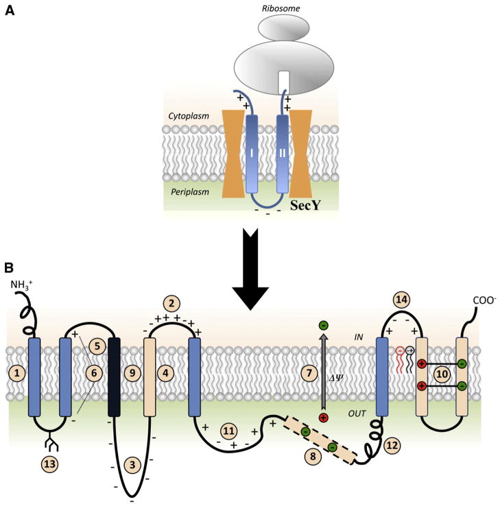
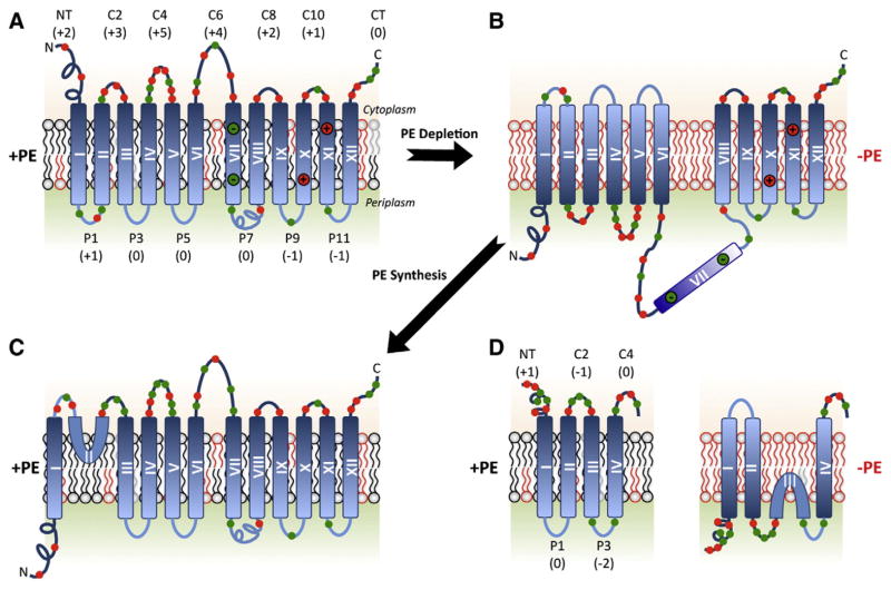
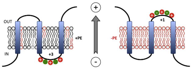
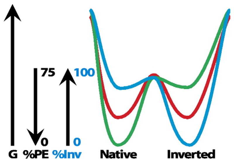
Membrane protein folding and topogenesis are tuned to a given lipid profile since lipids and proteins have co-evolved to follow a set of interdependent rules governing final protein topological organization. Transmembrane domain (TMD) topology is determined via a dynamic process in which topogenic signals in the nascent protein are recognized and interpreted initially by the translocon followed by a given lipid profile in accordance with the Positive Inside Rule. The net zero charged phospholipid phosphatidylethanolamine and other neutral lipids dampen the translocation potential of negatively charged residues in favor of the cytoplasmic retention potential of positively charged residues (Charge Balance Rule). This explains why positively charged residues are more potent topological signals than negatively charged residues. Dynamic changes in orientation of TMDs during or after membrane insertion are attributed to non-sequential cooperative and collective lipid-protein charge interactions as well as long-term interactions within a protein. The proportion of dual topological conformers of a membrane protein varies in a dose responsive manner with changes in the membrane lipid composition not only in vivo but also in vitro and therefore is determined by the membrane lipid composition. Switching between two opposite TMD topologies can occur in either direction in vivo and also in liposomes (designated as fliposomes) independent of any other cellular factors. Such lipid-dependent post-insertional reversibility of TMD orientation indicates a thermodynamically driven process that can occur at any time and in any cell membrane driven by changes in the lipid composition. This dynamic view of protein topological organization influenced by the lipid environment reveals previously unrecognized possibilities for cellular regulation and understanding of disease states resulting from mis-folded proteins. This article is part of a Special Issue entitled: Protein trafficking and secretion in bacteria. Guest Editors: Anastassios Economou and Ross Dalbey.

Keywords: Charge Balance Rule; Dual topology; Membrane protein topology; Phosphatidylethanolamine; Positive Inside Rule; Topogenesis.


Figures

Fig. 1
Fig. 1
Summary of factors determining membrane protein topogenesis and folding. (A) Initial orientation according to the Positive Inside Rule of a nascent two-TMD hairpin is shown in the SecY translocon channel. (B) The features of polytopic membrane proteins and biological membranes that influence folding and final TMD topology. TMDs are color coded for relative hydrophobicity: Light Blue indicates average hydrophobicity; Black indicates above average hydrophobicity; Beige indicates low hydrophobicity. Insertion efficiency of a TMD is driven by overall hydrophobicity with initial orientation determined within the translocon channel (#1) by the Positive Inside Rule (#2) or Charge Difference Rule (#6). Orientation and final topology are influenced by negatively charged residues present in high numbers (#3), flanking a marginally hydrophobic TMD (#4) or that lie at the end of a highly hydrophobic domain (#5) as well as the membrane potential (#7). After exit from the translocon into the lipid bilayer, topology is subject to interactions within the protein where TMDs of low hydrophobicity may become EMDs (#8), highly hydrophobic TMDs stabilize neighboring TMDs of low hydrophobicity (#9), or charged TMDs form salt bridges in the lipid bilayer (#10). Domains with conflicting signals can provide dynamic molecular hinges between independently folding domains (#8 and #11). Rapid, stable folding of an EMD (#12) or co-translational glycosylation of EMDs in eukaryotic cells (#13) can prevent transmembrane shuffling by the translocon. Finally, lipid–protein interactions dictated by the Charge Balance Rule (#14) determine topological organization during initial membrane insertion as well as after folding of membrane proteins.
Fig. 2
Fig. 2
Manipulating membrane phospholipid composition in vivo using E. coli. (A) Pathways native to E. coli are noted with blue arrows, and pathways resulting from foreign genes introduced into E. coli are noted with red arrows [50]. Lipids are color coded as zwitterionic (blue), neutral (green), anionic (orange) or cationic (gray). The genes encoding the following enzymes and associated with each biosynthetic step are listed next to the arrows: (1) CDP-diacylglycerol synthase; (2) PS synthase; (3) PS decarboxylase; (4) PGP synthase; (5) PGP phos-phatases; (6) CL synthases; (7) PG: membrane derived oligosaccharide sn-glycerol-1-P transferase; (8) diacylglycerol kinase; (9) GlcDAG synthase (Acholeplasma laidlawii); (10) GlcGlcDAG synthase (A. laidlawii); (11) PC synthase (Legionella pneumophila); (12) PI synthase (Saccharomyces cerevisiae); (13) N-Acyl PE synthase; (14) O-Acyl PG synthase; and (15) O-Lysyl PG synthase (Staphylococcus aureus) utilizing Lysyl-tRNA as the lysine donor. (B) Lipid profiles displayed by thin layer chromatography of 32PO4-labeled E. coli mutants with altered lipid compositions. Lane 1. AL95 (ΔpssA) has wild type phospholipid composition (80 mol% PE and 20 mol% PG plus CL) due to complementation by a plasmid (pDD72) copy of the pssA gene that encodes the committed step to PE biosynthesis. Lane 2. AL95 is PE-lacking due to the null allele of the pssA gene and contains mainly CL and PG. Lane 3. Introduction of the mgs gene from A. laidlawii into strain AL95 results in 35 mol% GlcDAG. The remaining lipids are primarily PG (35 mol%) and CL (25 mol%). Lane 4. Introduction into AL95 of the mgs and dgs genes from A. laidlawii results in about 30–40 mol% GlcGlcDAG with less than 1 mol% GlcDAG. Lane 5. Introduction into AL95 of the pcs gene from L. pneumophila results in about 70 mol% PC with the remainder being PG (26 mol%) plus CL (2.5 mol%). Lane 6. UE54 carries a null allele of the pgsA gene encoding the committed step to PG and CL biosynthesis making it devoid of PG and CL and containing about 90 mol% PE, 4 mol% PA and 3.2 mol% CDP-diacylglycerol. Lane 7. Introduction into AL95 of the mprF gene from S. aureus results in about 60 mol% of O-Lysyl-PG (LPG) with the remainder being PG (9 mol%) plus CL (30 mol%) and other minor lipids. Lane 8. Introduction of the mprF gene into “wild-type” AL95/pDD72 results in about 23 mol% of LPG, 44 mol% of PE, 11 mol% of PG and 22 mol% of CL.
Fig. 3
Fig. 3
Topological organization of LacY, PheP, and GabP as a function of membrane lipid composition. TMDs (Roman numerals) and EMDs (Arabic numerals) are sequentially numbered from the N-terminus to C-terminus with EMDs exposed to the periplasm (P) or cytoplasm (C) as in wild type cells. Net charge of EMDs and distribution of positive (red) and negative (green) charges are shown. Topology of LacY is shown after initial assembly in PE-containing cells (A), after either initial assembly in PE-lacking cells or after dilution of PE following initial assembly in PE-containing cells (B), or after initiation of PE synthesis post-assembly of LacY in PE-lacking cells (C). EMD P7 is recognized by monoclonal antibody 4B1 in PE-containing (specific conformation) but not in PE-lacking (loss of native conformation) cells. Charges in TMD VII salt bridge with charges in TMDs X and XI in native LacY. The interconversion of topological conformers (A, B and C) is reversible in both directions. Topology and EMD charge distribution of the lipid sensitive domains of PheP and GabP are shown in PE-containing and PE-lacking cells (D).
Fig. 4
Fig. 4
PE and the Charge Balance Rule. A cytoplasmic EMD is shown containing a mixture of negatively and positively charged amino acids. Left Panel. According to the Charge Balance Rule PE (black) would raise the pKa and suppress the translocation potential of negatively charged residues (green), which increases the effective positive charge potential of the EMD (+3) thus favoring its retention on the cytoplasmic side of the membrane. Right Panel. In the absence of PE (red) negatively charged residues exert their full translocation potential and result in translocation of the domain that now exhibits a lower effective net positive charge (+1). Even though the charge is still net +1, negatively charged residues may exhibit stronger signals than positively charged residues in the absence of PE. The membrane potential (positive outward) determines EMD directionality. This figure was modified from the original figure published in [85] © Annual Reviews of Biochemistry.
Fig. 5
Fig. 5
Dual minima energy folding funnel for LacY as a function of membrane lipid composition. The schematic depicts a two-dimensional section through the three-dimensional protein folding funnel. The folding of LacY to its lowest free energy state (G) proceeds via a funnel-shaped energy landscape whose shape is defined by the physicochemical properties of the lipid environment (green, 75% PE; red, intermediate % PE; blue, 0% PE). The topology of the N-terminal bundle of LacY is responsive to the membrane PE content. During initial folding of LacY multiple topological conformers are in rapid equilibrium, but subsequent folding events result in a stable mixture of conformers determined by the percent PE and separated by a high activation energy. A change in the PE content raises the free energy of the conformers resulting in a redistribution of conformers governed by the new PE content. This figure was originally published in [60] © the American Society for Biochemistry and Molecular Biology.

References

    1. Enquist K, Fransson M, Boekel C, Bengtsson I, Geiger K, Lang L, Pettersson A, Johansson S, von Heijne G, Nilsson I. Membrane-integration characteristics of two ABC transporters, CFTR and P-glycoprotein. J Mol Biol. 2009;387:1153–1164. - PubMed
    1. Lee E, Manoil C. Mutations eliminating the protein export function of a membrane-spanning sequence. J Biol Chem. 1994;269:28822–28828. - PubMed
    1. Hessa T, Kim H, Bihlmaier K, Lundin C, Boekel J, Andersson H, Nilsson I, White SH, von Heijne G. Recognition of transmembrane helices by the endoplasmic reticulum translocon. Nature. 2005;433:377–381. - PubMed
    1. Mackenzie KR. Folding and stability of alpha-helical integral membrane proteins. Chem Rev. 2006;106:1931–1977. - PubMed
    1. White SH, von Heijne G. Do protein–lipid interactions determine the recognition of transmembrane helices at the ER translocon? Biochem Soc Trans. 2005;33:1012–1015. - PubMed

Publication types

MeSH terms