

Case Reports
Treatment of renal angiomyolipoma: surgery versus angioembolization
- PMID: 24342162
- PMCID: PMC3926473
Item in Clipboard
Case Reports
Treatment of renal angiomyolipoma: surgery versus angioembolization
G Chir.
2013 Nov-Dec.
Abstract
Renal angiomyolipoma (AML) is a benign mesenchymal tumour. AML often leads to haemorrhagic complications such as retroperitoneal haematoma. Treatment varies from case to case, ranging from minimally invasive approaches such as selective embolization of the renal artery to invasive wedge resection, partial nephrectomy or, in more severe cases, radical nephrectomy. Here we report a case of retroperitoneal haematoma secondary to AML, treated with conservative approach by super-selective embolization of the lower-pole segmental renal artery.
Figures
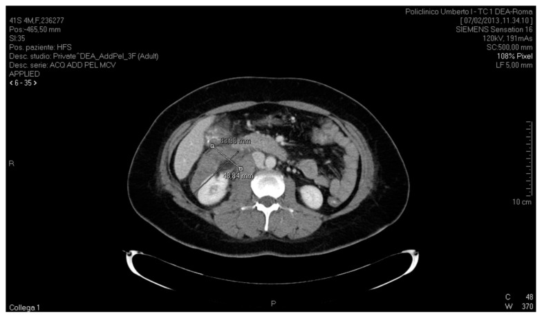
CT scan of the abdomen, axial section, showing a retroperitoneal serohaematic collection in the anterior peri-renal space and along the right parietal colic sulcus, with a diameter of 83.96 mm × 48.84 mm.
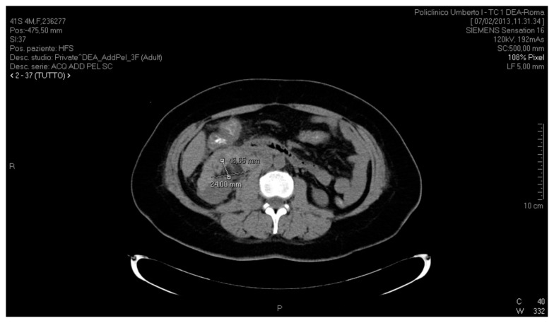
CT scan of the abdomen, axial section, showing a hypodense lesion anterior to the lower third of the kidney, with a maximum diameter of 46.65 mm × 24 mm.
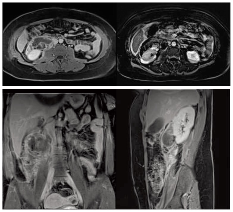
MRI scan; axial, sagittal and coronal sections showing a right retroperitoneal haematoma extending to the pelvis and a nodular exophytic, fat-containing lesion of the right kidney.
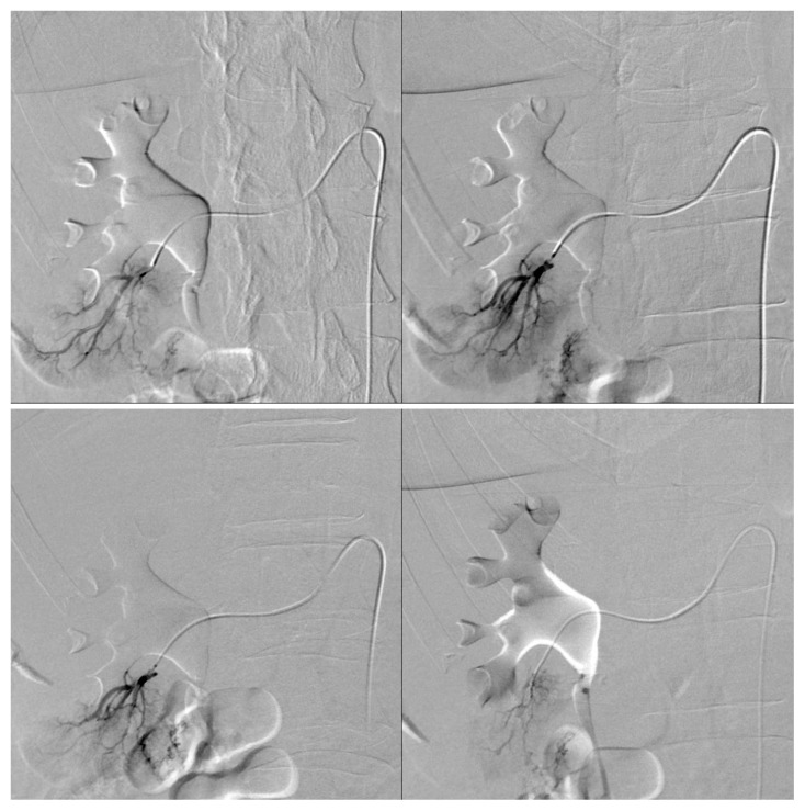
Pre-embolization angiogram showing two small arteries supplying the lesion after superselective catheterization of the lower-pole segmental renal artery.

Post-embolization angiogram showing cessation of blood flow in the branch of the right lower-pole segmental renal artery supplying the lesion.
References
-
- Fujii Y, Ajima J, Oka K, et al. Benign renal tumors detected among healthy adults by abdominal ultrasonography. Eur Urol. 1995;27:124–127. - PubMed
-
- Di Matteo G, Maturo A, Marzullo A, Peparini N, Wedard BM, Zeri KP, Di Matteo FM, Mascagni D. Giant abdominopelvic epithelioid angiomyolipoma associated with tuberous sclerosis: report of a case. Surg Today. 1999;29(11):1183–8. - PubMed
-
- Hadley DA, Bryant LJ, Ruckle HC. Conservative treatment of renal angiomyolipomas in patients with tuberous sclerosis. Clin Nephrol. 2006;65:22–7. - PubMed
Publication types
MeSH terms
LinkOut - more resources
Full Text Sources
Medical