Clinical Trial
. 2014 Feb;3(2):255-64.
doi: 10.5966/sctm.2013-0090. Epub 2013 Dec 16.

Pre- and postnatal transplantation of fetal mesenchymal stem cells in osteogenesis imperfecta: a two-center experience

Affiliations
Clinical Trial

Pre- and postnatal transplantation of fetal mesenchymal stem cells in osteogenesis imperfecta: a two-center experience

Cecilia Götherström et al. Stem Cells Transl Med. 2014 Feb.

Abstract

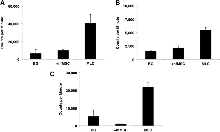
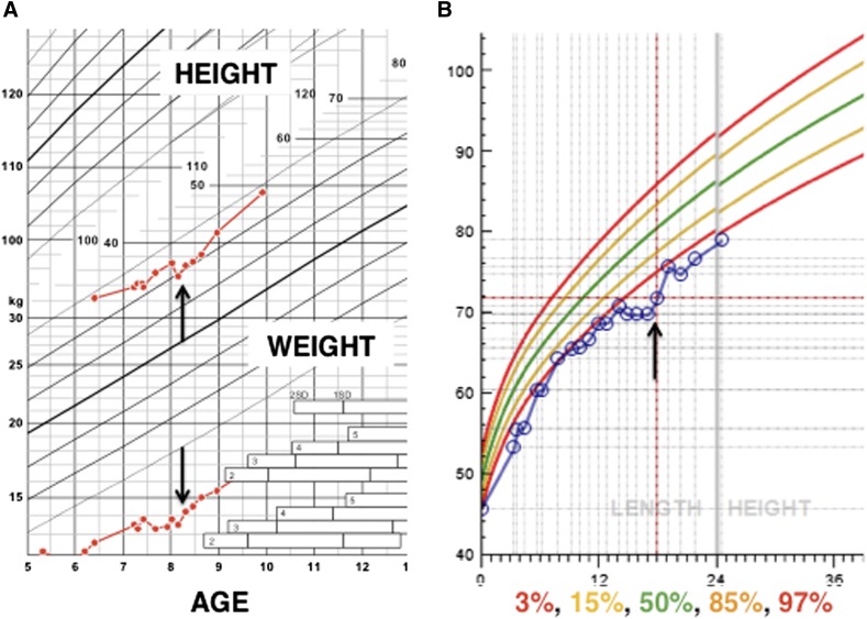
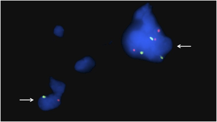
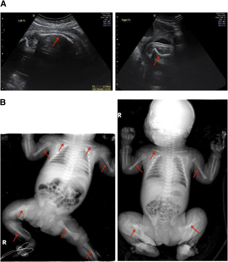
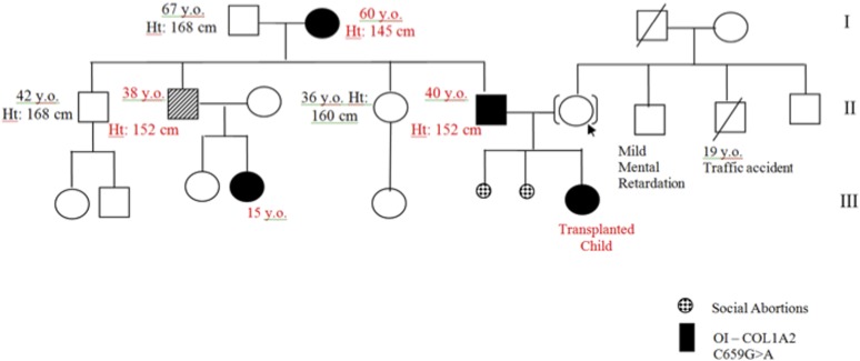
Osteogenesis imperfecta (OI) can be recognized prenatally with ultrasound. Transplantation of mesenchymal stem cells (MSCs) has the potential to ameliorate skeletal damage. We report the clinical course of two patients with OI who received prenatal human fetal MSC (hfMSC) transplantation and postnatal boosting with same-donor MSCs. We have previously reported on prenatal transplantation for OI type III. This patient was retransplanted with 2.8 × 10(6) same-donor MSCs per kilogram at 8 years of age, resulting in low-level engraftment in bone and improved linear growth, mobility, and fracture incidence. An infant with an identical mutation who did not receive MSC therapy succumbed at 5 months despite postnatal bisphosphonate therapy. A second fetus with OI type IV was also transplanted with 30 × 10(6) hfMSCs per kilogram at 31 weeks of gestation and did not suffer any new fractures for the remainder of the pregnancy or during infancy. The patient followed her normal growth velocity until 13 months of age, at which time longitudinal length plateaued. A postnatal infusion of 10 × 10(6) MSCs per kilogram from the same donor was performed at 19 months of age, resulting in resumption of her growth trajectory. Neither patient demonstrated alloreactivity toward the donor hfMSCs or manifested any evidence of toxicities after transplantation. Our findings suggest that prenatal transplantation of allogeneic hfMSCs in OI appears safe and is of likely clinical benefit and that retransplantation with same-donor cells is feasible. However, the limited experience to date means that it is not possible to be conclusive and that further studies are required.

Keywords: Cell therapy; In utero transplantation; Mesenchymal stem cells; Mesenchymal stromal cells; Osteogenesis imperfecta; Prenatal transplantation.


Figures

Figure 1.
Figure 1.
Immunological reaction toward donor cells. MLC was performed to evaluate patient A’s and patient B’s immunological reactions toward donor cells. Overall, 10,000 hfMSCs or 100,000 allogeneic peripheral blood lymphocytes were cocultured with 100,000 patient peripheral blood lymphocytes. (A): Patient A before postnatal transplantation, 8 years after prenatal transplantation. (B): Patient B before prenatal transplantation. (C): Patient B at birth, 7 weeks after prenatal transplantation. In all cases, an immune response was detected against allogeneic lymphocytes (MLC) but not against donor hfMSCs. Data are reported as mean ± SD of triplicate experiments. Abbreviations: BG, background; hfMSC, human fetal mesenchymal stem cell; MLC, mixed lymphocyte culture.
Figure 2.
Figure 2.
Growth curves for patients A and B. (A): Over the 2 years following postnatal transplantation (arrows), patient A’s linear growth improved from −6.5 to −6 standard deviations, and she is almost following her own growth velocity centiles. (B): Patient B’s growth followed a line just under the 3rd centile until 12 months of age, where it plateaued. Her growth resumed at just under the 3rd centile after postnatal transplantation of same-donor human fetal mesenchymal stem cells (arrow). Length is given in centimeters, weight in kilograms.
Figure 3.
Figure 3.
Detection of male cells in sections from a bone biopsy taken after postnatal transplantation. Fluorescence in situ hybridization analysis of 4-μm single sections for X and Y chromosomes using α-satellite probes for the centromeric regions. The bone sample was taken 9 months after postnatal transplantation at 8 years and 11 months of age from patient A. X chromosomes (red dots) and Y chromosomes (green dots) can be recognized in 4′,6-diamidino-2-phenylindole-stained cell nuclei (blue). In total, 4 Y-chromosome-positive cells were detected among 60,000 cells. The arrows indicate cells containing Y chromosomes. Original magnification, ×100.
Figure 4.
Figure 4.
Skeletal examination of patient B. (A): Fetal ultrasound examination at 26 weeks showed the presence of short long bones and multiple fresh healing and healed fractures (arrows). (B): A full-body radiological skeletal survey at birth confirmed the occurrence of healed fractures, indicated by arrows.
Figure 5.
Figure 5.
Pedigree tree of patient B’s family history of mutation indicating osteogenesis imperfecta. Genotyping of patient B’s family showed the same mutation in the patient’s father, first uncle, first cousin, and paternal grandmother, suggesting an autosomal dominant mode of inheritance. Abbreviations: Ht, height; y.o., years old.

Similar articles

Cited by

References

    1. Forlino A, Cabral WA, Barnes AM, et al. New perspectives on osteogenesis imperfecta. Nat Rev Endocrinol. 2011;7:540–557. - PMC - PubMed
    1. Steiner RD, Pepin MG, Byers PH. Osteogenesis Imperfecta. In: Pagon RA, Bird TD, Dolan CR et al., eds. GeneReviews. Seattle, WA: University of Washington, Seattle, 1993.
    1. Glorieux FH, Bishop NJ, Plotkin H, et al. Cyclic administration of pamidronate in children with severe osteogenesis imperfecta. N Engl J Med. 1998;339:947–952. - PubMed
    1. Bishop N. Characterising and treating osteogenesis imperfecta. Early Hum Dev. 2010;86:743–746. - PubMed
    1. Pittenger MF, Mackay AM, Beck SC, et al. Multilineage potential of adult human mesenchymal stem cells. Science. 1999;284:143–147. - PubMed

Publication types

Substances

Supplementary concepts