. 2014 May;28(5):1145-8.
doi: 10.1038/leu.2013.377. Epub 2013 Dec 17.

BCL2-specific inhibitor ABT-199 synergizes strongly with cytarabine against the early immature LOUCY cell line but not more-differentiated T-ALL cell lines

Affiliations

BCL2-specific inhibitor ABT-199 synergizes strongly with cytarabine against the early immature LOUCY cell line but not more-differentiated T-ALL cell lines

N M Anderson et al. Leukemia. 2014 May.
No abstract available


Conflict of interest statement

Conflict of interest

The authors declare no conflicts of interest.

Figures
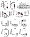
Figure 1
Figure 1. BCL2 and BCLw are upregulated in both primary early immature T-ALL and LOUCY cells, and BCL2 inhibition by ABT-263 or ABT-199 induces apoptosis in LOUCY cells
(a) BCL2 and BCLw transcripts are overexpressed in primary early immature T-ALL patient samples (n=9), compared to more differentiated T-ALL (n=46) cases. (b) Western blot analysis of BCL2, BCLw and BCL-XL in the early immature LOUCY cell line and seven more differentiated cell lines. JURKAT cells overexpressing BCL2 is used as a positive control for BCL2 and ACTIN serves as a loading control. (c-d) Among the eight T-ALL cell lines tested, LOUCY cells exhibit the most sensitivity to BCL2 inhibition by ABT-263 (c; IC50 = 43 nM) and ABT-199 (d; IC50 = 18 nM). Cell viability was determined with Cell Titer Blue (Promega) after 48 hours of treatment. (e,f) Annexin V and PI staining was used to determine apoptosis in LOUCY, ALL-SIL and JURKAT cells upon 48 hours of treatment with ABT-263 (e) and ABT-199 (f). Values in (a, c-f) are means ± SD, and represent three biological replicates. Statistical significance was determined by 2-tailed t-test, *, P<0.05; **, P<0.001; ***, P<0.0005.
Figure 2
Figure 2. Cytarabine synergizes with ABT-199 in chemoresistant LOUCY cell line
(a) Among the eight cell lines treated with cytarabine, the LOUCY cell line was the most resistant. (b,c) Combination treatment of ABT-199 and cytarabine depicted as normalized isobolograms shows synergy between the two drugs in LOUCY and JURKAT cell lines. Calcusyn software was used to analyze combination data to produce the isobolograms normalized to the IC50 of each drug. The black diagonal line connects x- and y-axes of the normalized isobologram and red dots on the black line represent additive dose combinations; red dots below the black line represent synergistic drug combinations and red dots above the black line represent antagonism. The LOUCY and JURKAT cell lines were treated with the following combination doses of ABT-199 and cytarabine for 7 days and 48 hours respectively. LOUCY: ABT-199 from 1.2 to 33nM and cytarabine from 0.4 to 11nM and JURKAT: ABT-199 from 0.078 to 5mM and cytarabine from 0.031 to 0.5mM. (d-e) Cell cycle analysis and apoptosis analysis was performed on LOUCY cells after 24 hours of treatment with DMSO, ABT-199 (15nM), cytarabine (78nM) and both drugs in combination (ABT-199: 15nM; cytarabine: 78nM). (d) Twenty-four hours after treatment LOUCY cells were fixed and stained with propidium iodide (PI), and cells were analyzed for Sub-G1, G0/G1, S and G2/M. (e) Apoptosis analysis was performed on cells stained with Annexin V and PI 24 hours after treatment. Cell viability was determined with Cell Titer Blue (Promega) after 48 hours of treatment. Values in (a, d, e) are means ± SD, and represent three biological replicates. Statistical significance was determined by 2-tailed t-test, **, P<0.005; ***, P<0.0007.

References

    1. Pui CH, Robison LL, Look AT. Acute lymphoblastic leukaemia. Lancet. 2008;371(9617):1030–43. - PubMed
    1. Goldberg JM, Silverman LB, Levy DE, Dalton VK, Gelber RD, Lehmann L, et al. Childhood T-cell acute lymphoblastic leukemia: the Dana-Farber Cancer Institute acute lymphoblastic leukemia consortium experience. Journal of clinical oncology : official journal of the American Society of Clinical Oncology. 2003;21(19):3616–22. - PubMed
    1. Coustan-Smith E, Mullighan CG, Onciu M, Behm FG, Raimondi SC, Pei D, et al. Early T-cell precursor leukaemia: a subtype of very high-risk acute lymphoblastic leukaemia. Lancet Oncol. 2009;10(2):147–56. - PMC - PubMed
    1. Van Vlierberghe P, Ambesi-Impiombato A, Perez-Garcia A, Haydu JE, Rigo I, Hadler M, et al. ETV6 mutations in early immature human T cell leukemias. J Exp Med. 2011;208(13):2571–9. - PMC - PubMed
    1. Rothenberg EV, Moore JE, Yui MA. Launching the T-cell-lineage developmental programme. Nature reviews Immunology. 2008;8(1):9–21. - PMC - PubMed

Publication types

MeSH terms