

Review
doi: 10.4161/auto.27367.
Epub 2013 Dec 9.
The vacuole versus the lysosome: when size matters
Affiliations
- PMID: 24343261
- PMCID: PMC5396098
- DOI: 10.4161/auto.27367
Item in Clipboard
Review
The vacuole versus the lysosome: when size matters
Autophagy.
2014 Feb.
Abstract
The morphometric examination of autophagic bodies provides useful information about the mechanism and magnitude of macroautophagy, and yeast researchers frequently utilize various measurements of these structures as part of their quantification of the process. The utility of this approach, however, has led to the common misconception that autophagic bodies can be found in the mammalian lysosome, which is generally not correct.
Keywords: autophagic body; autophagy; lysosome; stress; vacuole.
Figures
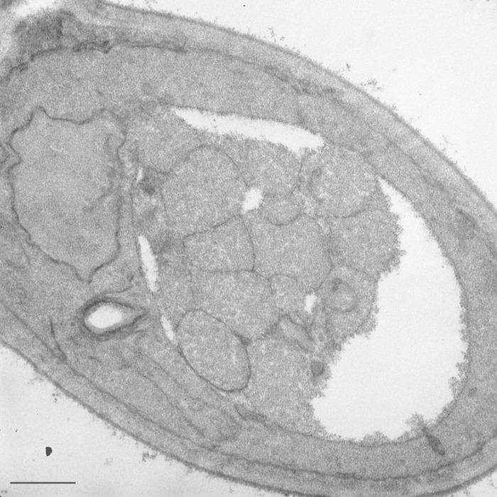
Figure 1. Autophagic bodies in the yeast vacuole. A yeast strain harboring a deletion of the PEP4 gene was grown in rich medium and shifted to nitrogen-starvation conditions for 4 h. Scale bar: 500 nm.
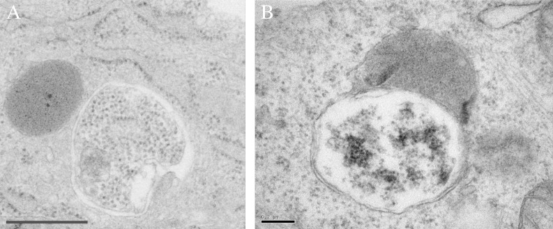
Figure 3. Mammalian lysosomes are too small to accommodate autophagic bodies. Mouse embryonic fibroblasts showing an autophagosome next to (A) or fusing with (B) an electron dense lysosome. Scale bars: 600 nm (A) and 200 nm (B).
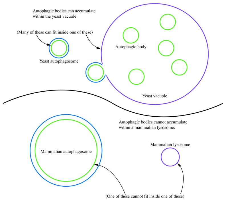
Figure 4. Autophagic bodies can accumulate within the yeast vacuole, but not the mammalian lysosome. In this schematic diagram, the relative sizes of yeast and mammalian autophagosomes are shown compared with the vacuole and lysosome. Even smaller mammalian autophagosomes are typically too large to allow the release of the inner vesicle into the lysosome lumen.
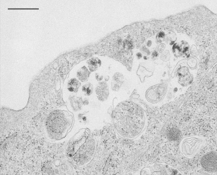
Figure 2. Single-membrane compartments in mouse embryonic fibroblasts. The “autophagic bodies” are likely generated by the fusion of an autophagosome with a late endosome/multivesicular body. This figure was reproduced from Figure 5B of Eskelinen, E-L. Meth Mol Biol, 2008; 445:11–28 with the permission of Springer.
References
-
- Indge KJ. . The isolation and properties of the yeast cell vacuole. J Gen Microbiol 1968; 51:441 - 6; http://dx.doi.org/10.1099/00221287-51-3-441; PMID: 4968622 - DOI - PubMed
-
- Matile P, Wiemken A. . The vacuole as the lysosome of the yeast cell. Arch Mikrobiol 1967; 56:148 - 55; http://dx.doi.org/10.1007/BF00408765; PMID: 4873367 - DOI - PubMed
-
- Pratt P, Bryce J, Stewart G. . The yeast vacuole—a scanning electron microscopy study during high gravity wort fermentations. J Inst Brew 2007; 113:55 - 60; http://dx.doi.org/10.1002/j.2050-0416.2007.tb00256.x - DOI
-
- Eskelinen E-L, Reggiori F, Baba M, Kovács AL, Seglen PO. . Seeing is believing: the impact of electron microscopy on autophagy research. Autophagy 2011; 7:935 - 56; http://dx.doi.org/10.4161/auto.7.9.15760; PMID: 21566462 - DOI - PubMed
-
- Dice JF, Klionsky DJ. . Artophagy: the art of autophagy--macroautophagy. Autophagy 2010; 6:320 - 1; http://dx.doi.org/10.4161/auto.6.3.11263; PMID: 20118653 - DOI - PubMed
Publication types
MeSH terms
Substances
Grants and funding
LinkOut - more resources
Full Text Sources
Other Literature Sources