Review
. 2014 Mar;35(3):515-27.
doi: 10.1093/carcin/bgt480. Epub 2013 Dec 16.

Cancer as a metabolic disease: implications for novel therapeutics

Affiliations
Review

Cancer as a metabolic disease: implications for novel therapeutics

Thomas N Seyfried et al. Carcinogenesis. 2014 Mar.

Abstract

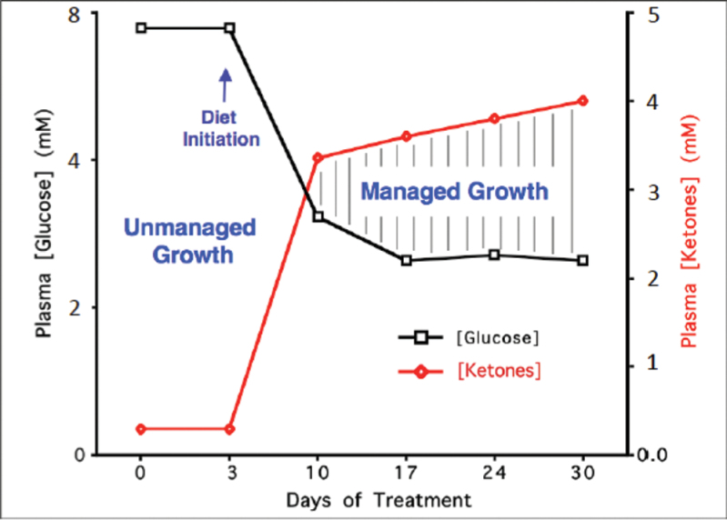
Emerging evidence indicates that cancer is primarily a metabolic disease involving disturbances in energy production through respiration and fermentation. The genomic instability observed in tumor cells and all other recognized hallmarks of cancer are considered downstream epiphenomena of the initial disturbance of cellular energy metabolism. The disturbances in tumor cell energy metabolism can be linked to abnormalities in the structure and function of the mitochondria. When viewed as a mitochondrial metabolic disease, the evolutionary theory of Lamarck can better explain cancer progression than can the evolutionary theory of Darwin. Cancer growth and progression can be managed following a whole body transition from fermentable metabolites, primarily glucose and glutamine, to respiratory metabolites, primarily ketone bodies. As each individual is a unique metabolic entity, personalization of metabolic therapy as a broad-based cancer treatment strategy will require fine-tuning to match the therapy to an individual's unique physiology.


Figures

Fig. 1.
Fig. 1.
Role of the nucleus and mitochondria in the origin of tumors. This image summarizes the experimental evidence supporting a dominant role of the mitochondria in the origin of tumorigenesis as described previously (49). Normal cells are depicted in green with mitochondrial and nuclear morphology indicative of normal respiration and nuclear gene expression, respectively. Tumor cells are depicted in red with abnormal mitochondrial and nuclear morphology indicative of abnormal respiration and genomic instability. (1) Normal cells beget normal cells. (2) Tumor cells beget tumor cells. (3) Delivery of a tumor cell nucleus into a normal cell cytoplasm begets normal cells despite the persistence of tumor-associated genomic abnormalities. (4) Delivery of a normal cell nucleus into a tumor cell cytoplasm begets tumor cells or dead cells but not normal cells. The results suggest that tumors do not arise from nuclear genomic defects alone and that normal mitochondria can suppress tumorigenesis. Original diagram from Jeffrey Ling and Thomas N. Seyfried, with permission.
Fig. 2.
Fig. 2.
Typical ultrastructure of a normal mitochondrion and a mitochondrion from a human glioblastoma. Normal mitochondria contain elaborate cristae, which are extensions of the inner membrane and contain the protein complexes of the electron transport chain necessary for producing ATP through OxPhos. The mitochondrion from the glioblastoma (m) is enlarged and shows a near total breakdown of cristae (cristolysis) and an electron-lucent matrix. The absence of cristae in glioblastoma mitochondria indicates that OxPhos would be deficient. The arrow indicates an inner membrane fold. Bar: 0.33 μm. Method of staining: uranyl acetate/lead citrate. The glioblastoma multiforme mitochondrion was reprinted with permission from Journal of Electron Microscopy (94). The normal mitochondrion diagram was from http://academic.brooklyn.cuny.edu/biology/bio4fv/page/mito.htm.
Fig. 3.
Fig. 3.
Mitochondrial respiratory dysfunction as the origin of cancer. Cancer can arise from any number of non-specific events that damage the respiratory capacity of cells over time. The path to carcinogenesis will occur only in those cells capable of enhancing energy production through fermentation (substrate level phosphorylation, SLP). Despite the shift from respiration to SLP the ΔG′ of ATP hydrolysis remains fairly constant at approximately −56 kJ indicating that the energy from SLP compensates for the reduced energy from OxPhos. The mitochondrial stress response or retrograde signaling will initiate oncogene upregulation and tumor suppressor gene inactivation that are necessary to maintain viability of incipient cancer cells when respiration becomes unable to maintain energy homeostasis. Genomic instability will arise as a secondary consequence of protracted mitochondrial stress from disturbances in the intracellular and extracellular microenvironment. Metastasis arises from respiratory damage in cells of myeloid/macrophage origin (146). The degree of malignancy is linked directly to the energy transition from OxPhos to SLP. This scenario links all major cancer hallmarks to an extrachromosomal respiratory dysfunction (141). The T signifies an arbitrary threshold when the shift from OxPhos to SLP might become irreversible. Reprinted with modifications from (17).
Fig. 4.
Fig. 4.
Timeline of events following expression of K-Ras. The time axis depicts the various events after stimulation of the K-RAS gene expression. The findings of Huang et al. indicate that mitochondria-linked changes are observed around the time of the increase in the K-Ras protein (142). This is then followed by other changes, such as alteration in cell metabolism. Gene mutations would form as a downstream epiphenomenon of altered metabolism. Malignant transformation, documented by the colony-forming activity of the cells and their propensity to form tumors ensue much later. According to these findings, the Warburg effect (aerobic fermentation) arises as a consequence of K-RAS-induced respiratory injury. This timeline is in general agreement with Warburg’s central theory and with other similar findings that respiratory disturbance is an initial event in K-RAS-induced tumorigenesis (237–239). The timeline will be greatly protracted in vivo as shown from the Roskelley et al. (240) experiments. Reprinted from Neuzil et al. (143) with permission.
Fig. 5.
Fig. 5.
Relationship of circulating levels of glucose and ketones (β-hydroxybutyrate) to tumor management. The glucose and ketone values are within normal physiological ranges under fasting conditions in humans and will produce anti-angiogenic, anti-inflammatory and pro-apoptotic effects. We refer to this state as the zone of metabolic management. Metabolic stress will be greater in tumor cells than in normal cells when the whole body enters the metabolic zone. The values for blood glucose in mg/dl can be estimated by multiplying the mM values by 18. The glucose and ketone levels predicted for tumor management in human cancer patients are 3.1–3.8mM (55–65mg/dl) and 2.5–7.0mM, respectively. These ketone levels are well below the levels associated with ketoacidosis (blood ketone values greater than 15 mmol). Elevated ketones will protect the brain from hypoglycemia. Modified from a previous version (241).
Fig. 6.
Fig. 6.
The KD and HBO2T are synergistic in reducing systemic metastatic cancer in the syngeneic VM mouse model. VM-M3/Fluc tumor cells were implanted subcutaneously and systemic organ metastasis was evaluated ex vivo using bioluminescent imaging as described previously (242,243). Tumor growth was slower in mice fed the KD than in mice fed a standard high carbohydrate diet. (A) Representative animals from each treatment group demonstrating tumor bioluminescence at day 21 after tumor cell inoculation. Treated animals showed less bioluminescence than controls with KD + HBO2T mice exhibiting a profound decrease in tumor bioluminescence compared with all groups. (B) Total body bioluminescence was measured weekly as a measure of tumor size; error bars represent standard error of the mean. KD + HBO2T mice exhibited significantly less tumor bioluminescence than control animals at week 3 (P < 0.01; two-tailed student’s t-test) and an overall trend of notably slower tumor growth than controls and other treated animals throughout the study. (C and D) Day 21 organ bioluminescence of standard high carbohydrate diet and KD + HBO2T animals (N = 8) demonstrated a trend of reduced metastatic tumor burden in animals receiving the combined therapy. Spleen bioluminescence was significantly decreased in KD + HBO2T mice (*P < 0.02; two-tailed student’s t-test). Reprinted with permission from Poff et al. (243).

References

    1. Sporn M.B. (1996). The war on cancer. Lancet, 347, 1377–1381 - PubMed
    1. Seyfried T.N. (2012). Cancer As a Metabolic Disease: On the Origin, Management, and Prevention of Cancer. John Wiley & Sons, Hoboken, NJ
    1. Fidler I.J. (2003). The pathogenesis of cancer metastasis: the ‘seed and soil’ hypothesis revisited. Nat. Rev. Cancer, 3, 453–458 - PubMed
    1. Lazebnik Y. (2010). What are the hallmarks of cancer? Nat. Rev. Cancer, 10, 232–233 - PubMed
    1. Tarin D. (2011). Cell and tissue interactions in carcinogenesis and metastasis and their clinical significance. Semin. Cancer Biol., 21, 72–82 - PubMed

Publication types