. 2014 Mar;82(3):1074-83.
doi: 10.1128/IAI.01028-13. Epub 2013 Dec 16.

Low-dose intradermal infection with trypanosoma congolense leads to expansion of regulatory T cells and enhanced susceptibility to reinfection

Affiliations

Low-dose intradermal infection with trypanosoma congolense leads to expansion of regulatory T cells and enhanced susceptibility to reinfection

Chukwunonso Onyilagha et al. Infect Immun. 2014 Mar.

Abstract

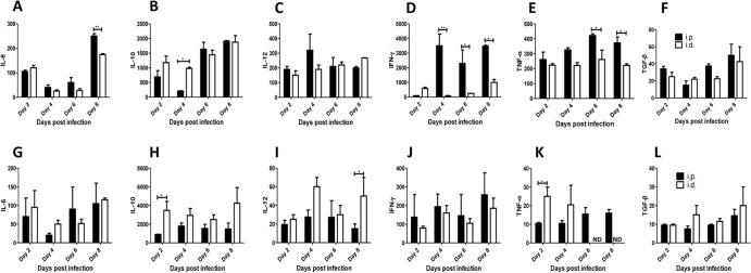
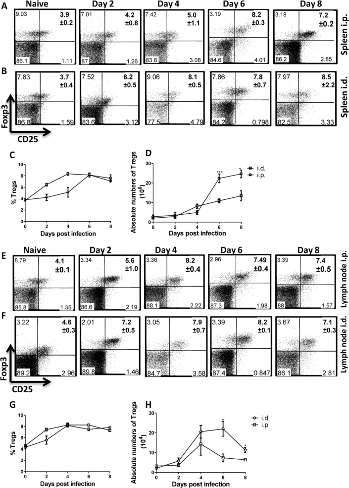
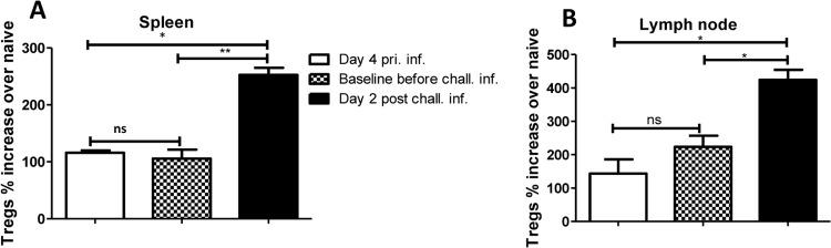
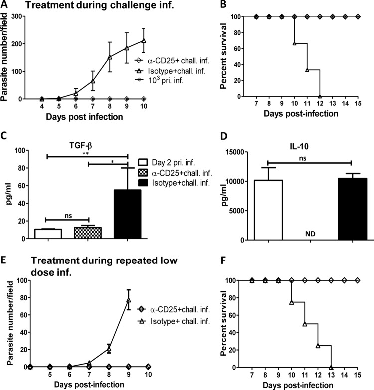
BALB/c mice are highly susceptible to experimental intraperitoneal Trypanosoma congolense infection. However, a recent report showed that these mice are relatively resistant to primary intradermal low-dose infection. Paradoxically, repeated low-dose intradermal infections predispose mice to enhanced susceptibility to an otherwise noninfectious dose challenge. Here, we explored the mechanisms responsible for this low-dose-induced susceptibility to subsequent low-dose challenge infection. We found that akin to intraperitoneal infection, low-dose intradermal infection led to production of interleukin-10 (IL-10), IL-6, IL-12, tumor necrosis factor alpha (TNF-α), transforming growth factor β (TGF-β), and gamma interferon (IFN-γ) by spleen and draining lymph node cells. Interestingly, despite the absence of parasitemia, low-dose intradermal infection led to expansion of CD4+ CD25+ Foxp3+ cells (T regulatory cells [Tregs]) in both the spleens and lymph nodes draining the infection site. Depletion of Tregs by anti-CD25 monoclonal antibody (MAb) treatment during primary infection or before challenge infection following repeated low-dose infection completely abolished the low-dose-induced enhanced susceptibility. In addition, Treg depletion was associated with dramatic reduction in serum levels of TGF-β and IL-10. Collectively, these findings show that low-dose intradermal infection leads to rapid expansion of Tregs, and these cells mediate enhanced susceptibility to subsequent infection.


Figures

FIG 1
FIG 1
Low-dose i.d. T. congolense infection and susceptibility to challenge infection. Groups of BALB/c mice were infected intraperitoneally or intradermally (4 mice per group) with 102 and 103 Trypanosoma congolense (clone TC13) parasites. At indicated time points, parasitemia (A) was monitored by microscopy by counting the number of parasites in the blood taken from the tail vein of infected mice. The survival of infected mice was also determined (B). Note that all mice infected i.d. with 102 and 103 TC13 parasites controlled the infection, while all mice infected i.p. with 102 and 103 parasites developed infection and died within 8 to 10 days postinfection. In another experiment, groups of mice were infected i.d. (C) with 102 T. congolense parasites once a week for 2 weeks (which does not lead to parasitemia). One week after the last parasite injection, mice were reinfected (challenged) intradermally with 103 T. congolense parasites. Four naive age-matched mice were also infected with 103 T. congolense parasites i.d. or i.p. as controls. Parasitemia (D) and survival period (E) were determined. The results presented are representative of 4 different experiments with similar results. Error bars show means ± SEM.
FIG 2
FIG 2
Cytokine profile in mice infected i.d. and i.p. with 103 T. congolense parasites. Groups of mice (4 to 5 per group) were infected either i.d. or i.p. with 103 T. congolense parasites, and at indicated times, the mice were sacrificed and the spleen and draining lymph node cells were cultured in complete medium in the presence of freeze-thawed T. congolense. After 72 h, the levels of IL-6, IL-10, IL-12, IFN-γ, TNF-α, and TGF-β in the cell culture supernatant fluids were determined by ELISA and expressed as change (pg/ml) over levels in cells from uninfected (naive) animals. Panels A to F represent cytokine levels in the spleen, and panels G to L represent cytokine levels in the lymph node. The data presented are representative of 3 different experiments with similar results. Error bars show means ± SEM. *, P < 0.05; **, P < 0.01. ND, not detected (i.e., below the ELISA sensitivity).
FIG 3
FIG 3
Primary low-dose intradermal infection-induced susceptibility is durable. BALB/c mice (n = 6) were infected (inf.) intradermally with 102 T. congolense parasites weekly (for 2 consecutive weeks [Fig. 1C]). After 30 days of no detectable parasitemia, some mice (n = 3) were treated three times with cyclophosphamide (at 3-day intervals), while others were challenged (i.d.) with 103 T. congolense parasites and monitored for parasitemia (A). In addition, blood samples from uninfected (naive) or infected mice (i.d. or i.p.) were assessed for the presence of T. congolense DNA by PCR. Parasites purified from the blood of i.p.-infected mice by diethylaminoethyl (DEAE)-cellulose anion-exchange chromatography were used as the positive control (B). In another experiment, BALB/c mice (n = 8) were infected (i.d.) twice with 102 T. congolense parasites (once a week for 2 weeks [Fig. 1C]). After 40 days of no detectable parasitemia, some mice (n = 4) and some age-matched controls (n = 3) were reinfected with 103 T. congolense parasites, and parasitemia (C) and survival (D) were monitored. chall. inf., challenge infection. The results presented are representative of 2 different experiments with similar results. Error bars show means ± SEM.
FIG 4
FIG 4
Low-dose intradermal T. congolense infection leads to systemic expansion of CD4+ CD25+ Foxp3+ cells (Tregs). Groups of mice (4 or 5 mice per group) were infected either i.d. or i.p. with 103 T. congolense parasites. At the indicated days, infected mice were sacrificed, and single-cell suspensions of the spleen and draining lymph nodes were stained with fluorochrome-conjugated antibodies against CD4, CD25, and Foxp3, and the percentages (A, B, C, E, F, and G) and absolute numbers (D and H) of Tregs were assessed by flow cytometry. Panels A, B, E, and F are representative dot plots (percentages and standard deviations [SD]), while panels C and G are line graphs of the means ± SEM of 4 or 5 mice per group. The data presented are representative of 3 different experiments with similar results. Bars show means ± SEM. *, P < 0.05; **, P < 0.01; ***, P < 0.001.
FIG 5
FIG 5
Expansion of Tregs following primary intradermal infection is sustained. Eight mice were infected intradermally with 102 T. congolense parasites weekly (for 2 consecutive weeks). One week after the last infection, some mice (n = 4) were sacrificed to determine the baseline levels of Tregs. The remaining mice (n = 4) and some age-matched naive controls were challenged (chall. inf.) intradermally with 103 T. congolense parasites and sacrificed 4 days postchallenge to determine the levels of Tregs. Shown are the percentages of CD4+ CD25+ Foxp3+ cells (Tregs) in the spleens (A) and the lymph nodes (B) draining the infection site, presented as percent fold increase over the level in naive (uninfected) mice. pri. inf., primary infection. The data presented are representative of 2 different experiments with similar results. Error bars show means ± SEM. *, P < 0.05; **, P < 0.01. ns, not significant.
FIG 6
FIG 6
Depletion of Tregs abolishes intradermal low-dose-enhanced susceptibility to T. congolense infection. Groups of mice (n = 4 or 5) were injected weekly with 102 T. congolense parasites or PBS (for 2 consecutive weeks). On the 3rd week, mice were treated with anti-CD25 MAb PC61 (100 μg/mouse) or the isotype-matched control MAb and rechallenged (chall. inf.) with 103 T. congolense parasites the next day. Daily parasitemia (A) and survival period (B) were monitored as described in Materials and Methods. At the humane endpoint, mice were sacrificed, and sera were assessed for TGF-β (C) and IL-10 (D) by ELISA. In another experiment, groups of mice were injected with anti-CD25 or isotype control MAb (n = 3 or 4 mice/group) at each point of the primary (pri. inf.) low-dose (102 parasites) T. congolense infection (once weekly for 2 weeks). On the 3rd week, the mice were challenged with 103 T. congolense parasites, and parasitemia (E) and survival period (F) were determined. The data presented are representative of 3 (A to D) and 2 (E and F) different experiments with similar results. Bars show means ± SEM. *, P < 0.05; **, P < 0.01. ns, not significant; ND, not detected (i.e., below the ELISA sensitivity).
FIG 7
FIG 7
Depletion of macrophages does not abolish primary low-dose i.d. resistance and susceptibility to rechallenge infection. Groups of BALB/c mice (n = 4) were treated with liposomal chlodronate (Chlod.) to deplete macrophages or with control liposome (Lip). After 24 h, all mice were infected i.d. with 102 T. congolense parasites and monitored for parasitemia (A). Three weeks after primary infection (pri. inf.) (without parasitemia), all mice were rechallenged (chall. inf.) with 103 T. congolense parasites, and parasitemia (A) and survival (B) were monitored. The results presented are representative of 2 different experiments with similar results. Error bars show means ± SEM.

References

    1. Simarro PP, Cecchi G, Paone M, Franco JR, Diarra A, Ruiz JA, Fevre EM, Courtin F, Mattioli RC, Jannin JG. 2010. The atlas of human African trypanosomiasis: a contribution to global mapping of neglected tropical diseases. Int. J. Health Geogr. 9:57. 10.1186/1476-072X-9-57 - DOI - PMC - PubMed
    1. Stuart K, Brun R, Croft S, Fairlamb A, Gurtler RE, McKerrow J, Reed S, Tarleton R. 2008. Kinetoplastids: related protozoan pathogens, different diseases. J. Clin. Invest. 118:1301–1310. 10.1172/JCI33945 - DOI - PMC - PubMed
    1. Brun R, Don R, Jacobs RT, Wang MZ, Barrett MP. 2011. Development of novel drugs for human African trypanosomiasis. Future Microbiol. 6:677–691. 10.2217/fmb.11.44 - DOI - PubMed
    1. Hursey BS. 2001. The programme against African trypanosomiasis: aims, objectives and achievements. Trends Parasitol. 17:2–3. 10.1016/S1471-4922(00)01851-1 - DOI - PubMed
    1. Kristjanson PM, Swallow BM, Rowlands GJ, Kruska RL, de Leeuw PN. 1999. Measuring the costs of African animal trypanosomosis: the potential benefits of control and returns to research. Agric. Syst. 59:79–98. 10.1016/S0308-521X(98)00086-9 - DOI

Publication types

MeSH terms

LinkOut - more resources