. 2014 Mar;82(3):949-59.
doi: 10.1128/IAI.01233-13. Epub 2013 Dec 16.

Rosetting Plasmodium falciparum-infected erythrocytes bind to human brain microvascular endothelial cells in vitro, demonstrating a dual adhesion phenotype mediated by distinct P. falciparum erythrocyte membrane protein 1 domains

Affiliations

Rosetting Plasmodium falciparum-infected erythrocytes bind to human brain microvascular endothelial cells in vitro, demonstrating a dual adhesion phenotype mediated by distinct P. falciparum erythrocyte membrane protein 1 domains

Yvonne Adams et al. Infect Immun. 2014 Mar.

Abstract

Adhesion interactions between Plasmodium falciparum-infected erythrocytes (IE) and human cells underlie the pathology of severe malaria. IE cytoadhere to microvascular endothelium or form rosettes with uninfected erythrocytes to survive in vivo by sequestering IE in the microvasculature and avoiding splenic clearance mechanisms. Both rosetting and cytoadherence are mediated by the parasite-derived IE surface protein family Plasmodium falciparum erythrocyte membrane protein 1 (PfEMP1). Rosetting and cytoadherence have been widely studied as separate entities; however, the ability of rosetting P. falciparum strains to cytoadhere has received little attention. Here, we show that IE of the IT/R29 strain expressing a rosette-mediating PfEMP1 variant (IT4var09) cytoadhere in vitro to a human brain microvascular endothelial cell line (HBEC-5i). Cytoadherence was inhibited by heparin and by treatment of HBEC-5i with heparinase III, suggesting that the endothelial receptors for IE binding are heparan sulfate proteoglycans. Antibodies to the N-terminal regions of the IT4var09 PfEMP1 variant (NTS-DBL1α and DBL2γ domains) specifically inhibited and reversed cytoadherence down to low concentrations (<10 μg/ml of total IgG). Surface plasmon resonance experiments showed that the NTS-DBLα and DBL2γ domains bind strongly to heparin, with half-maximal binding at a concentration of ∼0.5 μM in both cases. Therefore, cytoadherence of IT/R29 IE is distinct from rosetting, which is primarily mediated by NTS-DBL1α interactions with complement receptor 1. These data show that IT4var09-expressing parasites are capable of dual interactions with both endothelial cells and uninfected erythrocytes via distinct receptor-ligand interactions.


Figures
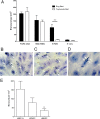
FIG 1
FIG 1
IT/R29 IE bind to HBEC-5i in ring-stage start assays. (A) Comparison of adhesion to HBEC-5i starting with pigmented trophozoite-stage-infected erythrocytes (IE) (white bars) or ring-stage IE (black bars) for parasite strains FCR3-CSA, HB3-HBEC, IT/R29, and IT-Uns. For the trophozoite-stage start assay, P. falciparum mature pigmented trophozoites at 5% parasitemia were incubated with HBEC-5i grown in 8-well chamber slides for 1 h before unbound IE were removed by gravity wash, fixed with 2% gluteraldehyde, and stained with Giemsa. For ring-stage start assay, IE at 5% parasitemia were coincubated overnight with HBEC-5i cells. The following day, unbound IE were removed by gravity wash and fixed and stained as described for the trophozoite-stage start assay. IE adhesion was assessed by microscopic examination of 10 fields (40× objective), and the mean numbers of IE bound and standard errors from four independent experiments are shown. The nonrosetting parasite strains FCR3-CSA, HB3-HBEC, and IT-Uns showed no difference in adhesion between ring-stage and pigmented trophozoite-stage start, whereas the rosetting IT/R29 strain showed significantly higher levels of adhesion after ring stage start (***, P < 0.005 by one-way ANOVA with Tukey's post hoc test). FCR3-CSA (B) and HB3-HBEC (C) adhesion to HBEC-5i show a diffuse scattering of adherent IE (white arrows). (D) IT/R29 adherent IE occur in clusters (white arrows). (E) Adhesion of IT/R29 IE to primary human pulmonary microvascular endothelial cells (HPMEC) and primary human brain microvascular endothelial cells (HBMEC) in ring-stage start assays as described for panels A and B. The mean numbers of IE bound and standard errors from three independent experiments are shown. Binding to HBMEC was significantly lower than that to HBEC-5i (**, P < 0.01 by one-way ANOVA with Tukey's post hoc test).
FIG 2
FIG 2
Antibodies against the IT4var09 PfEMP1 variant recognize IE bound to HBEC-5i. (A) Diagram of the IT4var09 PfEMP1 variant expressed by IT/R29 rosetting IE. The extracellular region is composed of multiple Duffy binding-like (DBL) and cysteine-rich interdomain regions (CIDR). TM, transmembrane region; ATS, acidic terminal segment. (B) Immunofluorescence assay (IFA) of IT/R29 IE adhering to HBEC-5i. Ring-stage IE at 5% parasitemia were incubated overnight with HBEC-5i in 8-well chamber slides. The following day the nonadherent IE were washed off and the slides fixed in acetone-methanol prior to immunostaining. Rabbit polyclonal antibodies against the IT4var09 NTS-DBL1α domain (anti-IT/R29) at 1:5,000 dilution were incubated for 1 h and then washed prior to detection with Alexa Fluor 488 goat anti-rabbit IgG at 1/1,000 dilution (green). The nuclei of the cells were stained with 1 μg/ml 4,6-diamidino-2-phenylindole (DAPI; blue; large nuclei are HBEC-5i, small nuclei are IE). Negative controls were IgG from a nonimmunized rabbit (rabbit IgG) and rabbit polyclonal antibodies to the NTS-DBL1α domain of an irrelevant PfEMP1 variant TM180var1 (anti-TM180) tested at 1/5,000 dilution. Slides were viewed with a 100× objective using a Leica DM LB2 fluorescence microscope, and images were taken at consistent exposure for all antibodies. Adherent IE staining with the IT4var09 antibodies are shown by white arrows.
FIG 3
FIG 3
IT4var09 antibodies inhibit and reverse IT/R29 IE adhesion to HBEC-5i. (A) Antibodies against IT4var09 domains at 100 μg/ml inhibited adhesion of IT/R29 IE to HBEC-5i, whereas negative-control antibodies (nonimmunized rabbit IgG and TM180var1 antibodies) did not. (B) Antibodies against IT4var09 domains were tested at 100 μg/ml to determine their ability to reverse adhesion of IT/R29 IE to HBEC-5i. Only NTS-DBL1α and DBL2γ antibodies were capable of significantly reversing adhesion. NTS-DBL1α and DBL2γ antibodies showed a dose-dependent effect in both inhibition (C) and reversal (D) experiments. Data shown are the means and standard errors from three independent experiments in all cases. Statistical significance was determined by one-way ANOVA with Tukey's post hoc test. ***, P < 0.005.
FIG 4
FIG 4
Adhesion of IT/R29 IE to HBEC-5i is heparan sulfate sensitive. (A) The ability of sulfated glycoconjugate compounds to reverse IT/R29 IE adhesion to HBEC-5i was tested by incubating cocultured IT/R29 IE and HBEC-5i for 1 h with 100 μg/ml of compound. Slides were then washed by gravity, fixed with 2% glutaraldehyde, stained with Giemsa, and assessed by microscopy (10 fields, 40× objective). The sulfated glycoconjugate compounds were dissolved in PBS, and the negative control was the addition of an equivalent volume of PBS alone with no added compound. (B) The ability of 100 μg/ml of heparin and heparan sulfate to reverse IT/R29 IE adhesion was performed as described for panel A. The effect of heparinase III on adhesion reversal was studied by incubation of cells with 0.2 IU/ml of enzyme for 2 h to remove heparan sulfate residues. Cells were washed, stained, and assessed by microscopy as described for panel A. Data shown are the means and standard errors from at least three independent experiments for panels A and B. (C) Soluble CSA (100 μg/ml for 2 h) and chondroitinase ABC enzyme (0.5 IU/ml for 2 h) were tested for their ability to reverse IT/R29 adhesion as described for panels A and B. (D) Heparinase III (0.2 IU/ml for 2 h) and chondroitinase ABC (0.5 IU/ml for 2 h) were tested for their effect on HB3-HBC adhesion. For panels C and D, the negative control was addition of an equivalent volume of PBS alone with no added compound/enzyme, and in each case means and standard errors from 2 independent experiments are shown. Statistical significance was determined by one-way ANOVA with Tukey's post hoc test. ***, P < 0.005.
FIG 5
FIG 5
IT4var09 NTS-DBL1α and DBL2γ domains bind heparin, and the binding is inhibited by sulfated glycoconjugates. (A) SPR signal for the binding of IT4var09 NTS-DBL1α from a maximum concentration of 4 μM to a heparin-coated surface. (B) SPR signal for the binding of DBL2γ from a maximum concentration of 4 μM to a heparin-coated surface. (C) Competition experiments in which the IT4var09 DBL2γ domain was incubated with different concentrations of glycoconjugates before assessment of binding to a heparin-coated surface.
FIG 6
FIG 6
Adhesion of IT/R29 IE to HBEC-5i is not CR1 dependent. Mouse monoclonal antibodies to CR1 were tested for their ability to inhibit adhesion of IT/R29 IE to HBEC-5i, and no significant inhibition was seen. Data shown are the means and standard errors from two independent experiments.

References

    1. Rowe JA, Claessens A, Corrigan RA, Arman M. 2009. Adhesion of Plasmodium falciparum-infected erythrocytes to human cells: molecular mechanisms and therapeutic implications. Expert Rev. Mol. Med. 11:e16. 10.1017/S1462399409001082 - DOI - PMC - PubMed
    1. Baruch DI, Pasloske BL, Singh HB, Bi X, Ma XC, Feldman M, Taraschi TF, Howard RJ. 1995. Cloning the P. falciparum gene encoding PfEMP1, a malarial variant antigen and adherence receptor on the surface of parasitised human erythrocytes. Cell 82:77–87. 10.1016/0092-8674(95)90054-3 - DOI - PubMed
    1. Su X-Z, Heatwole VM, Wertheimer SP, Guinet F, Herrfeldt JA, Peterson DS, Ravetch JA, Wellems TE. 1995. A large and diverse family gene family (var) encodes 200–350 kD proteins implicated in the antigenic variation and cytoadherence of Plasmodium falciparum-infected erythrocytes. Cell 82:89–99. 10.1016/0092-8674(95)90055-1 - DOI - PubMed
    1. Smith JD, Chitnis CE, Craig AG, Roberts DJ, Hudson-Taylor DE, Peterson DS, Pinches R, Newbold CI, Miller LH. 1995. Switches in expression of Plasmodium falciparum var genes correlate with changes in antigenic and cytoadherent phenotypes of infected erythrocytes. Cell 82:101–110. 10.1016/0092-8674(95)90056-X - DOI - PMC - PubMed
    1. Kraemer SM, Smith JD. 2006. A family affair: var genes, PfEMP1 binding, and malaria disease. Curr. Opin. Microbiol. 9:374–380. 10.1016/j.mib.2006.06.006 - DOI - PubMed

Publication types

MeSH terms

Substances

LinkOut - more resources