

Expression of six drug transporters in vaginal, cervical, and colorectal tissues: Implications for drug disposition in HIV prevention
- PMID: 24343710
- PMCID: PMC4061289
- DOI: 10.1002/jcph.248
Expression of six drug transporters in vaginal, cervical, and colorectal tissues: Implications for drug disposition in HIV prevention
Abstract
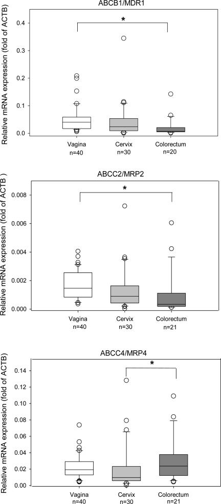
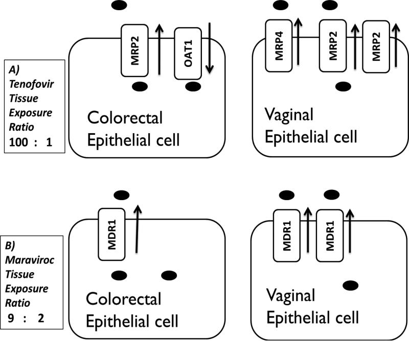
Effective antiretroviral (ARV)-based HIV prevention strategies require optimizing drug exposure in mucosal tissues; yet factors influencing mucosal tissue disposition remain unknown. We hypothesized drug transporter expression in vaginal, cervical, and colorectal tissues is a contributing factor and selected 3 efflux (ABCB1/MDR1, ABCC2/MRP2, ABCC4/MRP4) and 3 uptake (SLC22A6/OAT1, SLC22A8/OAT3, SLCO1B1/OATP1B1) transporters to further investigate based on their affinity for 2 ARVs central to prevention (tenofovir, maraviroc). Tissue was collected from 98 donors. mRNA and protein expression were quantified using qPCR and immunohistochemistry (IHC). Hundred percent of tissues expressed efflux transporter mRNA. IHC localized them to the epithelium and/or submucosa. Multivariable analysis adjusted for age, smoking, and co-medications revealed significant (P < 0.05) differences in efflux transporter mRNA between tissue types (vaginal ABCB1 3.9-fold > colorectal; vaginal ABCC2 2.9-fold > colorectal; colorectal ABCC4 2.0-fold > cervical). In contrast, uptake transporter mRNA was expressed in <25% of tissues. OAT1 protein was detected in 0% of female genital tissues and in 100% of colorectal tissues, but only in rare epithelial cells. These data support clinical findings of higher maraviroc and tenofovir concentrations in rectal tissue compared to vaginal or cervical tissue after oral dosing. Quantifying mucosal transporter expression and localization can facilitate ARV selection to target these tissues.
Keywords: HIV; antiretrovirals; mucosal; pharmacokinetics; prevention; transporters.
© 2014, The American College of Clinical Pharmacology.
Figures

Similar articles
-
Expression of Genes for Drug Transporters in the Human Female Genital Tract and Modulatory Effect of Antiretroviral Drugs.PLoS One. 2015 Jun 23;10(6):e0131405. doi: 10.1371/journal.pone.0131405. eCollection 2015. PLoS One. 2015. PMID: 26102284 Free PMC article.
-
In Vivo Modulation of Cervicovaginal Drug Transporters and Tissue Distribution by Film-Released Tenofovir and Darunavir for Topical Prevention of HIV-1.Mol Pharm. 2020 Mar 2;17(3):852-864. doi: 10.1021/acs.molpharmaceut.9b01121. Epub 2020 Feb 20. Mol Pharm. 2020. PMID: 32017579
-
Expression, regulation, and function of drug transporters in cervicovaginal tissues of a mouse model used for microbicide testing.Biochem Pharmacol. 2016 Sep 15;116:162-75. doi: 10.1016/j.bcp.2016.07.009. Epub 2016 Jul 21. Biochem Pharmacol. 2016. PMID: 27453435 Free PMC article.
-
Drug transporters in tissues and cells relevant to sexual transmission of HIV: Implications for drug delivery.J Control Release. 2015 Dec 10;219:681-696. doi: 10.1016/j.jconrel.2015.08.018. Epub 2015 Aug 13. J Control Release. 2015. PMID: 26278511 Free PMC article. Review.
-
Antiretroviral pharmacology in mucosal tissues.J Acquir Immune Defic Syndr. 2013 Jul;63 Suppl 2(0 2):S240-7. doi: 10.1097/QAI.0b013e3182986ff8. J Acquir Immune Defic Syndr. 2013. PMID: 23764642 Free PMC article. Review.
Cited by
-
Phase 1 Pharmacokinetic Trial of 2 Intravaginal Rings Containing Different Dose Strengths of Vicriviroc (MK-4176) and MK-2048.Clin Infect Dis. 2019 Mar 19;68(7):1129-1135. doi: 10.1093/cid/ciy652. Clin Infect Dis. 2019. PMID: 30289444 Free PMC article. Clinical Trial.
-
Short communication: cheminformatics analysis to identify predictors of antiviral drug penetration into the female genital tract.AIDS Res Hum Retroviruses. 2014 Nov;30(11):1058-64. doi: 10.1089/AID.2013.0254. Epub 2014 Mar 13. AIDS Res Hum Retroviruses. 2014. PMID: 24512359 Free PMC article.
-
HIV pre-exposure prophylaxis: mucosal tissue drug distribution of RT inhibitor Tenofovir and entry inhibitor Maraviroc in a humanized mouse model.Virology. 2014 Sep;464-465:253-263. doi: 10.1016/j.virol.2014.07.008. Epub 2014 Aug 7. Virology. 2014. PMID: 25105490 Free PMC article.
-
Pharmacokinetics of antiretrovirals in genital secretions and anatomic sites of HIV transmission: implications for HIV prevention.Clin Pharmacokinet. 2014 Jul;53(7):611-24. doi: 10.1007/s40262-014-0148-z. Clin Pharmacokinet. 2014. PMID: 24859035 Free PMC article. Review.
-
Drug transporter mRNA expression and genital inflammation in South African women on oral pre-exposure prophylaxis (PrEP).AIDS Res Ther. 2025 Feb 15;22(1):18. doi: 10.1186/s12981-025-00713-z. AIDS Res Ther. 2025. PMID: 39955595 Free PMC article.
References
-
- UNAIDS [26 September. 2013];UNAIDS report on the global AIDS epidemic. http://www.unaids.org/en/media/unaids/contentassets/documents/epidemiolo.... Published 20 November 2012.
-
- van der Straten A, Van Damme L, Haberer JE, Bangsberg DR. Unraveling the divergent results of pre-exposure prophylaxis trials for HIV prevention. Aids. 2012;26(7):F13–9. - PubMed
Publication types
MeSH terms
Substances
Grants and funding
LinkOut - more resources
Full Text Sources
Other Literature Sources
Medical
