. 2013 Dec 23;203(6):929-42.
doi: 10.1083/jcb.201306073.

Higher-order unfolding of satellite heterochromatin is a consistent and early event in cell senescence

Higher-order unfolding of satellite heterochromatin is a consistent and early event in cell senescence

Eric C Swanson et al. J Cell Biol. .

Abstract

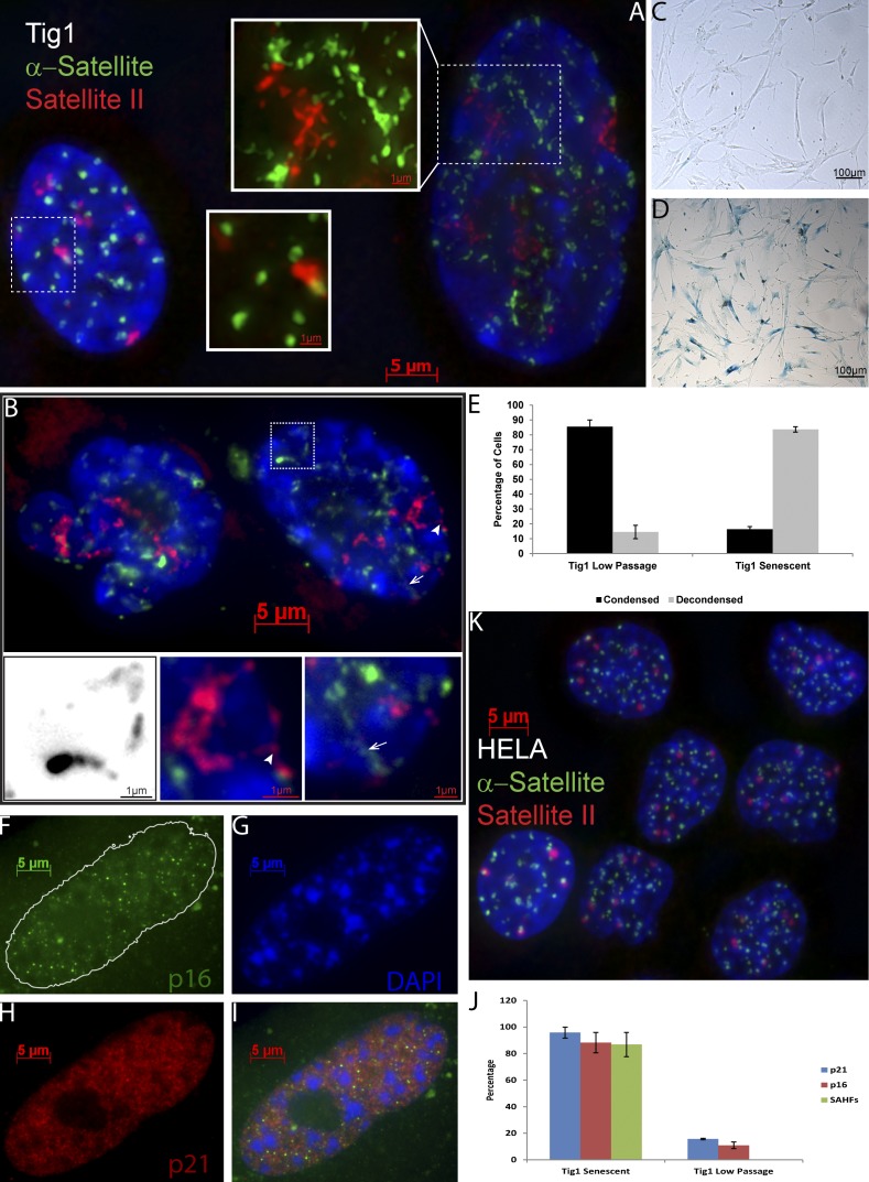
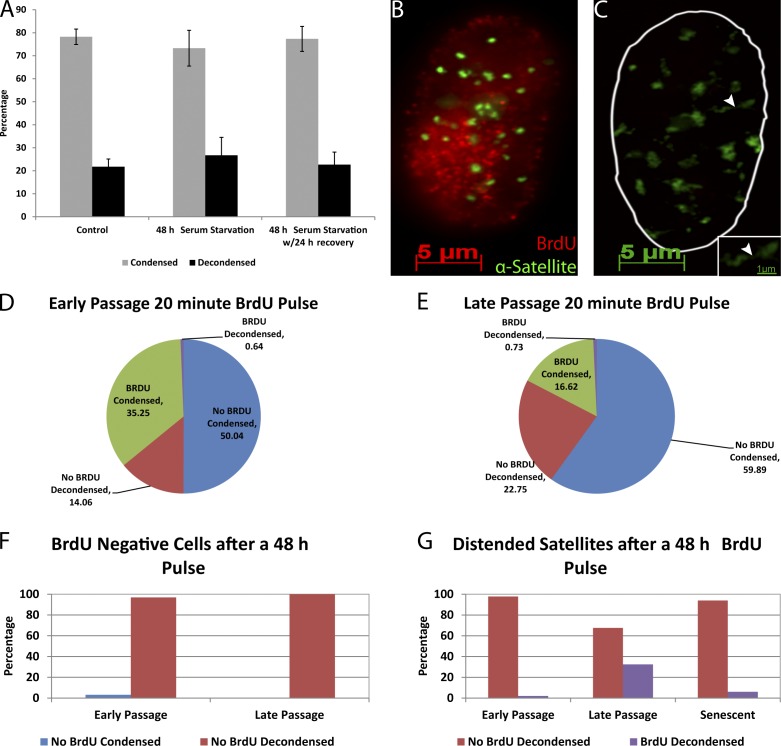
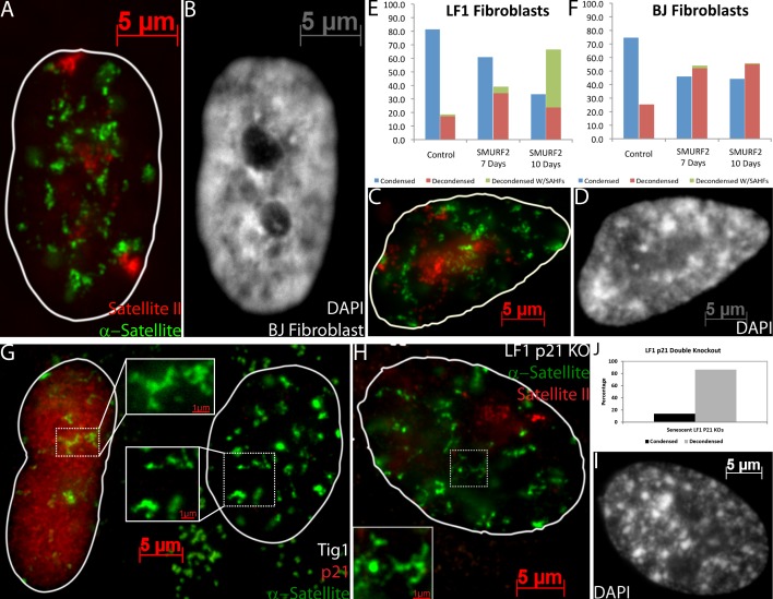
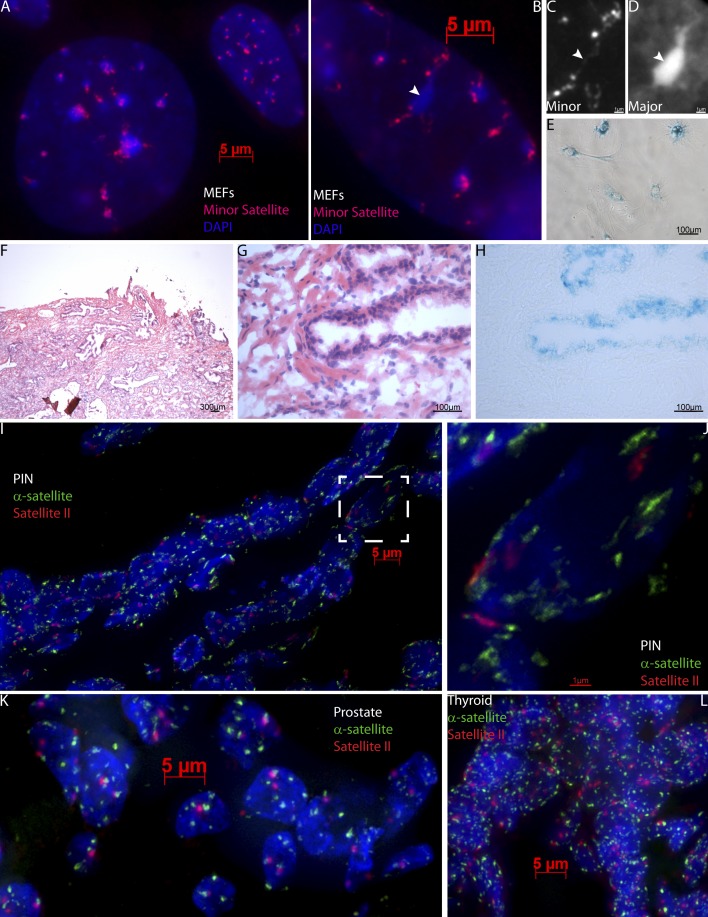
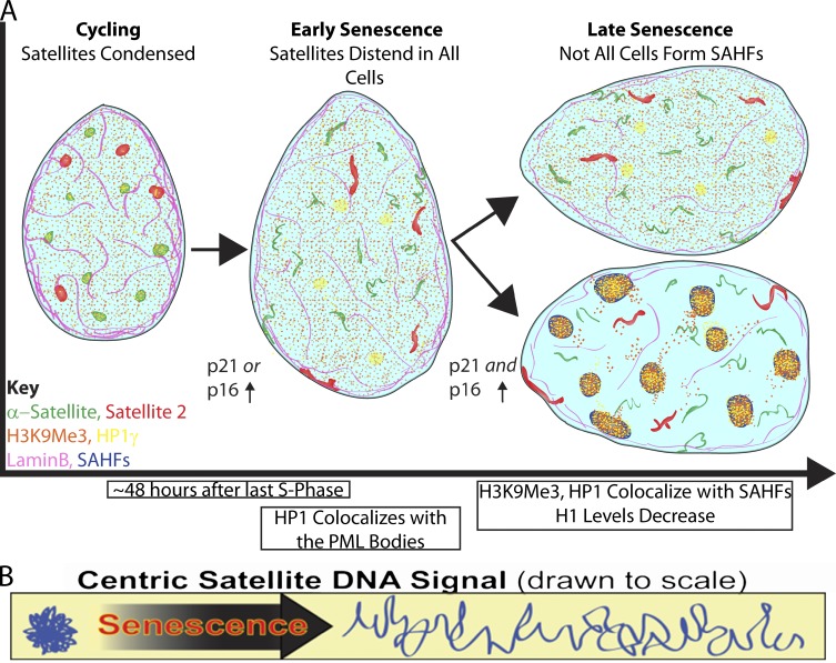
Epigenetic changes to chromatin are thought to be essential to cell senescence, which is key to tumorigenesis and aging. Although many studies focus on heterochromatin gain, this work demonstrates large-scale unraveling of peri/centromeric satellites, which occurs in all models of human and mouse senescence examined. This was not seen in cancer cells, except in a benign senescent tumor in vivo. Senescence-associated distension of satellites (SADS) occurs earlier and more consistently than heterochromatin foci formation, and SADS is not exclusive to either the p16 or p21 pathways. Because Hutchinson Guilford progeria syndrome patient cells do not form excess heterochromatin, the question remained whether or not proliferative arrest in this aging syndrome involved distinct epigenetic mechanisms. Here, we show that SADS provides a unifying event in both progeria and normal senescence. Additionally, SADS represents a novel, cytological-scale unfolding of chromatin, which is not concomitant with change to several canonical histone marks nor a result of DNA hypomethylation. Rather, SADS is likely mediated by changes to higher-order nuclear structural proteins, such as LaminB1.


Figures

Figure 1.
Figure 1.
Satellite DNA signals markedly distend in senescent fibroblasts. (A) α-Sat (green) and sat II (red) hybridization in early passage culture. Distended satellites are found in a small percentage of nuclei (right; see 3D images and Videos 1 and 2). (B) Most cells in late passage senescent cultures show distended satellites. Note that these cells do not have enlarged nuclei, but contain SAHF (DAPI). Insets show examples of threadlike satellite extensions, some of which extend to and contact the nuclear membrane (indicated by arrows and arrowheads). Inset of boxed region shown in black and white to enhance contrast. (C and D) β-Gal staining confirms that the cycling culture (C) contains few senescent cells and the senescent culture (D) contains many. (E) Quantification showing that senescent cultures contain more cells with distended satellites than cycling cultures. (F–J) Senescence was confirmed by staining for p16 (F), p21 (H), and SAHF (DAPI; G), and quantified (J; error bars are standard error). (K) HeLa cultures do not contain cells with distended satellites.
Figure 2.
Figure 2.
BrdU labeling demonstrates that distended satellites are characteristic of and specific to senescent fibroblasts. (A) Satellites do not distend in quiescent, serum-starved cells. (B and C) BrdU (red) labels cells with condensed satellites (green; B) but not cells with decondensed satellites (C). Inset shows high magnification of a distended satellite (arrowhead; cells are from same field). (D and E) Quantification of satellite distension in early (D) and late (E) passage cultures subjected to a 20-min BrdU pulse, confirming that cells with decondensed satellites are rarely in S phase. (F) Almost all cells that did not incorporate BrdU during a 48-h pulse had distended satellites (n = 100–200, from one of three repeats). (G) Early passage and senescent cells with distended satellites rarely incorporate BrdU during the previous 48-h period (n = 100–200, representative of one of three repeats). In presenescent late passage cultures in which more cells are entering senescence, a significant fraction of cells with distended satellites incorporated BrdU (error bars represent standard error).
Figure 3.
Figure 3.
SADS is a property common to multiple different senescence models and pathways. (A and B) SADS is present in a SMURF2-induced senescent BJ fibroblast, yet these cells do not form SAHF (B). (C and D) Oxidative stress-induced senescence of a Tig1 fibroblast results in distension of α-sat (green) and sat II (red) in a cell with SAHF (DAPI; D). (E and F) Quantification of SADS and SAHF in LF1 and BJ fibroblasts, induced with wild-type SMURF2 (n = 200, representative of one of three repeats). (G) Tig1 fibroblasts with SADS (green) may or may not be positive for p21 staining (red). (H–J) SADS in LF1 p21 knockout cells are quantified (n = 204; J) and a SAHF-positive senescent cell is shown (H and I) with α-sat (green), sat II (red), and DAPI (I).
Figure 4.
Figure 4.
SADS occur in MEFs and senescent cells within human tumors. (A) Cycling MEFs (top right) have condensed minor satellite (red), whereas senescent MEFs (bottom left) have distended minor satellite. (B–D) A senescent MEF with coalesced and elongated chromocenters (DAPI) and stringy minor satellite (red). The arrowhead points to a particularly elongated chromocenter (D) associated with a highly decondensed minor satellite (C). (E) Senescent MEFs, as shown in B, stain positive for β-gal. (F–J) Serial sections reveal SADS in PIN tissue. Using H&E staining (F and magnified in G) to identify PIN, which contains senescent cells as indicated by positive β-gal staining (H) and distended satellites by α-sat (green) and sat II (red; I). Signals from I (white box) are enlarged in J. (K and L) Malignant prostate (K) and thyroid (L) tumors, which lack senescent cells, typically have round, compact signals of α-sat (green) and sat II (red).
Figure 5.
Figure 5.
SADS is characteristic of HGPS cells that undergo premature arrest. (A) Frequency of distended satellites in early passage cells from two HGPS patients (1-yr-old HGADFN155 and 8-yr-old HGADFN167) and the older healthy mother of HGADFN167, HGMDFM090 (n = 100 in one of three representative experiments). (B) A 20-min BrdU pulse demonstrates that very few early passage HGADFN167 cells with decondensed satellites label, confirming that cells with SADS are rarely in S phase (n = 202). (C) Almost all cells that did not incorporate BrdU during a 48-h label had distended satellites (n = 100). (D–F) DNA FISH to α-sat (green) and sat II (red) shows that some HGPS cells have normal appearing compact satellites (E), whereas senescent cells in both the HGADFN167 (D) and HGADFN155 (F) cell lines contain SADS. Also notice the blebbed nuclear membrane (DAPI; F) characteristic of laminopathies. Arrows and arrowheads indicate a particularly distended α-sat signal that is enlarged in the corresponding insets.
Figure 6.
Figure 6.
Loss of compaction is specific to satellite DNA and is not caused by increased nuclear size. (A and B) DNA FISH to Chr. 17 α-sat (green) shows compact satellite signals in a cycling cell (A) and distended satellites in a senescent cell (B) as determined by the presence of SAHF (DAPI). The size of the corresponding signal is shown above the brackets. (C) The mean length of Chr. 17 α-sat in senescent cells is six times the mean length in cycling cells. (D) The shortest Chr. 17 α-sat measured in senescent cells is longer than the largest satellite measured in cycling cells. (E–H) Differences in satellite length cannot be explained by the twofold increase in nuclear diameter (E). The mean size of the Chr. 21 BAC signal (red) did not change (F) in cycling (G) or senescent (H) cells. (I–L) Telomere signal size (red) in cycling (I and J) or SMURF2-induced senescent (K and L) LF1 cells did not change. Senescence is judged by distension of the α-sat (green) and presence of SAHF (DAPI; error bars represent standard error).
Figure 7.
Figure 7.
Analysis of expression, heterochromatin proteins, DNA methylation, and histone modifications in satellites. (A and B) FISH to sat II RNA (green) shows no increased expression with SADS formation in senescent (left) versus cycling (right) cells, with satellite chromatin marked by CENP-B staining (red). (C) CENP-A (green) and CENP-B (red) are still present in senescent cells as determined by the presence of SAHF (DAPI) and decondensed CENP-B staining (magnified in inset). (D) Comparison of H3K9me3, H3K27me3, and H3K4me3 read densities between cycling and senescent IMR90 cells and between cycling IMR90 and H1hES cells on α-sat (RepeatMasker class: HSATII) sequences with corresponding Pearson’s r values. (E and F) As all cancer samples examined, U2OS cells have compact satellites (F; α-sat, green; sat II, red) and do not form SADS unless treated with 5-AzaC, which induces senescence (E). (G) Quantification of mean 1q12 signal areas (n > 60) in different cell samples (error bars represent standard error). (H) A DNA methylation-sensitive Southern blot with U2OS and cycling Tig1 cells digested by BstB1 (B), HpaII (H), or MspI (M) and detected with a probe for sat II 1q12 sequences. The arrow points to a band size that is lacking in some of the methylation-sensitive HpaII lanes, indicating that the 1q12 region is methylated in the corresponding cell types. (I–K) Cells with normal levels of LaminB1 (red; top left) rarely contain distended α-sat (green; I and K), but in cells with SADS (bottom right) LaminB1 is often diminished (J and K; n = 100 from one of three replicates).
Figure 8.
Figure 8.
Satellite distension in relation to other events in the cell senescence process. (A) In nuclei of cycling cells, the peri/centromeric satellite DNA signals are tightly compacted, but dramatically distend in senescent cells. This occurs before and irrespective of the later formation of SAHF, and in some cells within 48 h of the last S phase. SADS is seen in essentially all noncycling cells and occurs in cells that first up-regulate either the p21 or p16 pathway. In late stage senescence, both pathways are up-regulated, and the p16 pathway promotes formation of SAHF. During this process there is a reorganization of several heterochromatin factors as seen by immunofluorescence including H3K9Me3 and HP1γ, whereas H1 levels uniformly decrease with the formation of SAHF. Whereas nuclear diameter increases progressively and is most pronounced in cells with SAHF, SADS are seen in some cells before nuclear enlargement. The nuclear lamina protein (LaminB1) may play a role in the higher-order folding of the DNA as it is diminished in most cells before SADS form. (B) Sat DNA in cycling cells is tightly packaged and this packaging is lost during senescence as the DNA distends. Because three canonical histone modifications remain unchanged during senescence the difference in DNA packaging may be attributed to changes in higher-order folding.

References

    1. Adams P.D. 2007. Remodeling of chromatin structure in senescent cells and its potential impact on tumor suppression and aging. Gene. 397:84–93 10.1016/j.gene.2007.04.020 - DOI - PMC - PubMed
    1. Chandra T., Kirschner K., Thuret J.-Y., Pope B.D., Ryba T., Newman S., Ahmed K., Samarajiwa S.A., Salama R., Carroll T., et al. 2012. Independence of repressive histone marks and chromatin compaction during senescent heterochromatic layer formation. Mol. Cell. 47:203–214 10.1016/j.molcel.2012.06.010 - DOI - PMC - PubMed
    1. Chen Z., Trotman L.C., Shaffer D., Lin H.-K., Dotan Z.A., Niki M., Koutcher J.A., Scher H.I., Ludwig T., Gerald W., et al. 2005. Crucial role of p53-dependent cellular senescence in suppression of Pten-deficient tumorigenesis. Nature. 436:725–730 10.1038/nature03918 - DOI - PMC - PubMed
    1. Cristofalo V.J. 2005. SA beta Gal staining: biomarker or delusion. Exp. Gerontol. 40:836–838 10.1016/j.exger.2005.08.005 - DOI - PubMed
    1. De Cecco M., Criscione S.W., Peckham E.J., Hillenmeyer S., Hamm E.A., Manivannan J., Peterson A.L., Kreiling J.A., Neretti N., Sedivy J.M. 2013. Genomes of replicatively senescent cells undergo global epigenetic changes leading to gene silencing and activation of transposable elements. Aging Cell. 12:247–256 10.1111/acel.12047 - DOI - PMC - PubMed

Publication types