. 2014 Jan 7;111(1):39-44.
doi: 10.1073/pnas.1312833110. Epub 2013 Dec 16.

Initial stages of calcium uptake and mineral deposition in sea urchin embryos

Affiliations

Initial stages of calcium uptake and mineral deposition in sea urchin embryos

Netta Vidavsky et al. Proc Natl Acad Sci U S A. .

Abstract

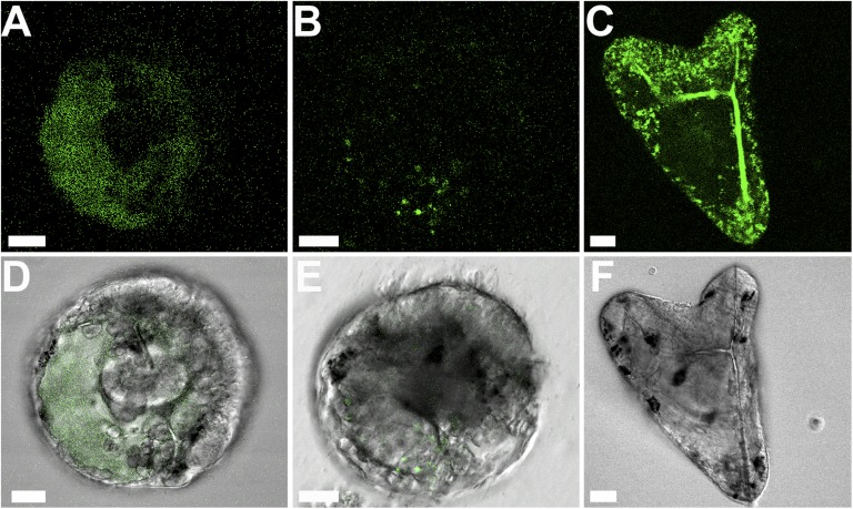
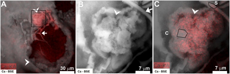
Sea urchin larvae have an endoskeleton consisting of two calcitic spicules. We reconstructed various stages of the formation pathway of calcium carbonate from calcium ions in sea water to mineral deposition and integration into the forming spicules. Monitoring calcium uptake with the fluorescent dye calcein shows that calcium ions first penetrate the embryo and later are deposited intracellularly. Surprisingly, calcium carbonate deposits are distributed widely all over the embryo, including in the primary mesenchyme cells and in the surface epithelial cells. Using cryo-SEM, we show that the intracellular calcium carbonate deposits are contained in vesicles of diameter 0.5-1.5 μm. Using the newly developed airSEM, which allows direct correlation between fluorescence and energy dispersive spectroscopy, we confirmed the presence of solid calcium carbonate in the vesicles. This mineral phase appears as aggregates of 20-30-nm nanospheres, consistent with amorphous calcium carbonate. The aggregates finally are introduced into the spicule compartment, where they integrate into the growing spicule.

Keywords: biomineralization; intracellular mineral deposition; mineralization pathway; sea urchin embryonic spicule; transient precursor mineral phase.


Conflict of interest statement

The authors declare no conflict of interest.

Figures

Fig. 1.
Fig. 1.
Cryo-SEM micrograph of a high-pressure frozen and freeze-fractured sea urchin embryo at the late gastrula stage. (A) The spicule cross-sections are marked with arrows. The archenteron (Ar) and epithelial cells (Ep) of the embryo are fractured, showing large numbers of intracellular vesicles of 0.5–1.5 μm. Note the multivesicular body adjacent to the PMC (arrowhead), enlarged in Fig. S1C. The blastocoel is marked with an asterisk. (B) The spicule cross-section (S; BSE image of the spicule area in Fig. S1B) is adjacent to a PMC that contains many vesicles, some empty and some packed with nanospheres (arrow). (C) Enlargement of the marked vesicle in B, showing nanospheres of 20–30 nm.
Fig. 2.
Fig. 2.
Cryo-SEM micrograph of a high-pressure frozen and freeze-fractured sea urchin embryo at the gastrula stage showing the spicule (S) and adjacent mineral-bearing vesicles, packed with nanospheres (arrowheads). BSE image in Fig. S3.
Fig. 3.
Fig. 3.
Confocal micrographs of live calcein-labeled sea urchin embryos at the gastrula stage. (AC) Green calcein fluorescence emission. (D and E) The fluorescence image in A and B is merged with the bright-field image. (F) Bright-field image of C. (A and D) The embryo received a 10-min calcein pulse at 40 h post fertilization (hpf), followed by a 1-h chase period in sea water. Calcein appears as a cloud in the blastocoel of the embryo and is not detected in an intracellular environment. One focal plane. (B and E) The embryo received a calcein pulse of 40 min at 40 hpf, followed by a 1-h chase period in sea water. Four focal planes, 2 μm apart, were stacked together. The calcein label is observed in the cellular environment in a more concentrated manner, in both the epithelial and other cells. (C and F) Embryo that was developed continuously in calcein-labeled sea water (46 hpf). Micrometer-size calcein-labeled granules are observed all over the embryo. One focal plane. Scale bars: 20 μm.
Fig. 4.
Fig. 4.
Cryo-SEM micrograph of a high-pressure frozen and freeze-fractured sea urchin embryo at the late gastrula stage. (A) The PMCs and epithelial cells (Ep) of the embryo are fractured, showing large numbers of intracellular vesicles (arrows). N, nuclei; S, spicule. (B) Epithelial cells, enlarged from A, showing fractured intracellular vesicles (arrow). (C and D) Fractured vesicle from an epithelial cell containing nanospheres (C) with corresponding BSE contrast image (D).
Fig. 5.
Fig. 5.
Correlative airSEM image of a ≤100-μm slice of a fixed embryo at ambient conditions showing the BSE signal (A), calcium EDS map (B), and calcein fluorescence (C). (D) The correlation between the calcium EDS signal (B) and calcein fluorescence (C) is shown in the superimposition of the two images. Yellow, colocalized signals; green and red, fluorescence and calcium EDS signals, appearing separately. Slight deformation of the surface, resulting from electron beam damage, may have decreased the actual colocalization area.
Fig. 6.
Fig. 6.
BSE image (white) and calcium EDS map (red) of a fixed and sliced embryo taken with the airSEM. (A) Whole embryo, with calcium EDS map (red) and BSE signal (white) superimposed. The spicule is marked with an arrow, and the membrane enveloping the embryo is marked with arrowheads. (B) Enlargement of the square-labeled area in A, containing a part of the spicule (arrow) and cell group (arrowhead); BSE image. (C) The same region as in B, with calcium EDS map (red) superimposed. EDS quantitative analysis of the marked areas is presented in Table 1. C, cell group; S, spicule.

References

    1. Volk GM, Goss LJ, Franceschi VR. Calcium channels are involved in calcium oxalate crystal formation in specialized cells of Pistia stratiotes L. Ann Bot (Lond) 2004;93(6):741–753. - PMC - PubMed
    1. Mahamid J, et al. Mapping amorphous calcium phosphate transformation into crystalline mineral from the cell to the bone in zebrafish fin rays. Proc Natl Acad Sci USA. 2010;107(14):6316–6321. - PMC - PubMed
    1. Lowenstam HA, Weiner S. Transformation of amorphous calcium phosphate to crystalline dahillite in the radular teeth of chitons. Science. 1985;227(4682):51–53. - PubMed
    1. Addadi L, Raz S, Weiner S. Taking advantage of disorder: Amorphous calcium carbonate and its roles in biomineralization. Adv Mater. 2003;15(12):959–970.
    1. Nakano E, Okazaki K, Iwamatsu T. Accumulation of radioactive calcium in larvae of the sea urchin Pseudocentrotus depressus. Biol Bull. 1963;125(1):125–132.

Publication types

LinkOut - more resources