

Microfluidics-assisted fabrication of gelatin-silica core-shell microgels for injectable tissue constructs
- PMID: 24344625
- PMCID: PMC3922064
- DOI: 10.1021/bm401533y
Microfluidics-assisted fabrication of gelatin-silica core-shell microgels for injectable tissue constructs
Abstract
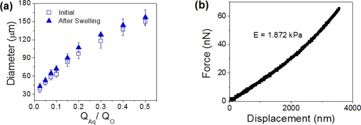
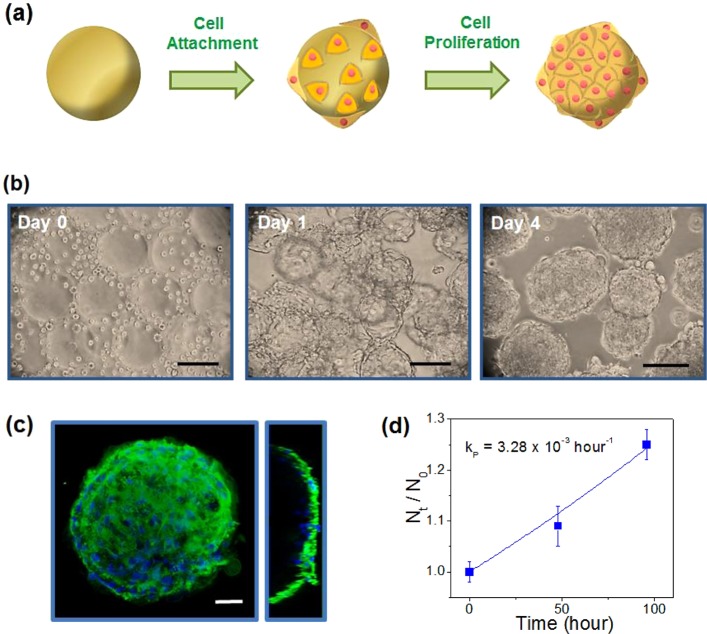
Microfabrication technology provides a highly versatile platform for engineering hydrogels used in biomedical applications with high-resolution control and injectability. Herein, we present a strategy of microfluidics-assisted fabrication photo-cross-linkable gelatin microgels, coupled with providing protective silica hydrogel layer on the microgel surface to ultimately generate gelatin-silica core-shell microgels for applications as in vitro cell culture platform and injectable tissue constructs. A microfluidic device having flow-focusing channel geometry was utilized to generate droplets containing methacrylated gelatin (GelMA), followed by a photo-cross-linking step to synthesize GelMA microgels. The size of the microgels could easily be controlled by varying the ratio of flow rates of aqueous and oil phases. Then, the GelMA microgels were used as in vitro cell culture platform to grow cardiac side population cells on the microgel surface. The cells readily adhered on the microgel surface and proliferated over time while maintaining high viability (∼90%). The cells on the microgels were also able to migrate to their surrounding area. In addition, the microgels eventually degraded over time. These results demonstrate that cell-seeded GelMA microgels have a great potential as injectable tissue constructs. Furthermore, we demonstrated that coating the cells on GelMA microgels with biocompatible and biodegradable silica hydrogels via sol-gel method provided significant protection against oxidative stress which is often encountered during and after injection into host tissues, and detrimental to the cells. Overall, the microfluidic approach to generate cell-adhesive microgel core, coupled with silica hydrogels as a protective shell, will be highly useful as a cell culture platform to generate a wide range of injectable tissue constructs.
Figures


References
-
- Hoffman A. S. Adv. Drug Delivery Rev. 2002, 54, 3–12. - PubMed
-
- Drury J. L.; Mooney D. J. Biomaterials 2003, 24, 4337–4351. - PubMed
-
- Peppas N. A.; Hilt J. Z.; Khademhosseini A.; Langer R. Adv. Mater. 2006, 18, 1345–1360.
-
- Burdick J. A.; Anseth K. S. Biomaterials 2002, 23, 4315–4323. - PubMed
-
- Seliktar D.; Zisch A. H.; Lutolf M. P.; Wrana J. L.; Hubbell J. A. J. Biomed. Mater. Res., Part A 2004, 68, 704–716. - PubMed
Publication types
MeSH terms
Substances
Grants and funding
- R01 EB012597/EB/NIBIB NIH HHS/United States
- HL092836/HL/NHLBI NIH HHS/United States
- EB012597/EB/NIBIB NIH HHS/United States
- DE019024/DE/NIDCR NIH HHS/United States
- R01 HL092836/HL/NHLBI NIH HHS/United States
- EB008392/EB/NIBIB NIH HHS/United States
- DE021468/DE/NIDCR NIH HHS/United States
- R01 DE021468/DE/NIDCR NIH HHS/United States
- HL099073/HL/NHLBI NIH HHS/United States
- R01 AR057837/AR/NIAMS NIH HHS/United States
- R01 HL099073/HL/NHLBI NIH HHS/United States
- AR057837/AR/NIAMS NIH HHS/United States
- RL1 DE019024/DE/NIDCR NIH HHS/United States
- R01 EB008392/EB/NIBIB NIH HHS/United States
LinkOut - more resources
Full Text Sources
Other Literature Sources
