Review
. 2014 Jan;24(1):83-97.
doi: 10.1111/bpa.12099.

The pathology of an autoimmune astrocytopathy: lessons learned from neuromyelitis optica

Affiliations
Review

The pathology of an autoimmune astrocytopathy: lessons learned from neuromyelitis optica

Claudia F Lucchinetti et al. Brain Pathol. 2014 Jan.

Abstract

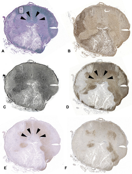
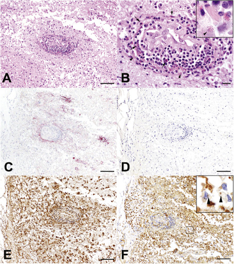
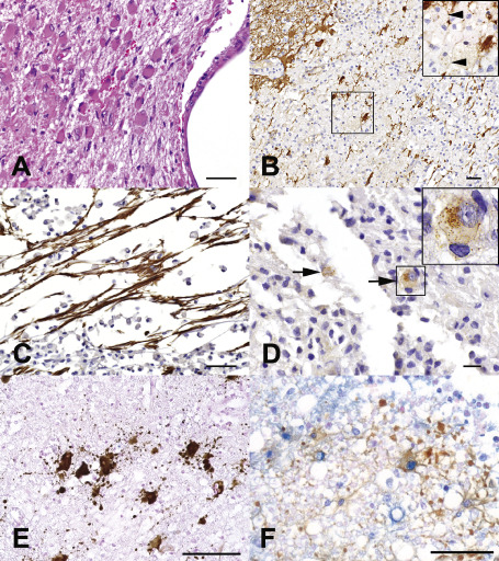
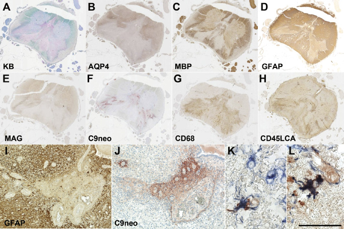
Neuromyelitis optica (NMO) is a disabling autoimmune astrocytopathy characterized by typically severe and recurrent attacks of optic neuritis and longitudinally extensive myelitis. Until recently, NMO was considered an acute aggressive variant of multiple sclerosis (MS), despite the fact that early studies postulated that NMO and MS may be two distinct diseases with a common clinical picture. With the discovery of a highly specific serum autoantibody (NMO-IgG), Lennon and colleagues provided the first unequivocal evidence distinguishing NMO from MS and other central nervous system (CNS) inflammatory demyelinating disorders. The target antigen of NMO-IgG was confirmed to be aquaporin-4 (AQP4), the most abundant water channel protein in the CNS, mainly expressed on astrocytic foot processes at the blood-brain barrier, subpial and subependymal regions. Pathological studies demonstrated that astrocytes were selectively targeted in NMO as evidenced by the extensive loss of immunoreactivities for the astrocytic proteins, AQP4 and glial fibrillary acidic protein (GFAP), as well as perivascular deposition of immunoglobulins and activation of complement even within lesions with a relative preservation of myelin. In support of these pathological findings, GFAP levels in the cerebrospinal fluid (CSF) during acute NMO exacerbations were found to be remarkably elevated in contrast to MS where CSF-GFAP levels did not substantially differ from controls. Additionally, recent experimental studies showed that AQP4 antibody is pathogenic, resulting in selective astrocyte destruction and dysfunction in vitro, ex vivo and in vivo. These findings strongly suggest that NMO is an autoimmune astrocytopathy where damage to astrocytes exceeds both myelin and neuronal damage. This chapter will review recent neuropathological studies that have provided novel insights into the pathogenic mechanisms, cellular targets, as well as the spectrum of tissue damage in NMO.

Keywords: aquaporin-4 (AQP4); astrocytopathy; neuromyelitis optica (NMO).


Figures

Figure 1
Figure 1
NMO spinal cord lesion. (A) Spinal cord cross‐section demonstrates extensive demyelination involving both the gray and white matter (LFB/PAS). (B) Extensive macrophage/microglia infiltration is present in the lesion (Kim1p). (C) Severe axonal loss is present in some areas of the lesion (Bielschowsky's staining). The lesion shows obvious loss of GFAP (D), AQP4 (E) and the glutamate transporter, EAAT2 (F). AQP4 loss is also evident in an area of the central spinal cord (E, arrowheads) where demyelination (A) and GFAP loss (D) are absent.
Figure 2
Figure 2
Perivascular NMO spinal cord lesion (boxed region in Figure  1). (A) A focal NMO lesion shows perivascular inflammation and tissue destruction in (H/E). (B) Lymphocytes and numerous perivascular eosinophils (arrows) are present, with evidence of eosinophil degranulation (arrowhead; H/E). (C) Complement C9neo deposition (C9neo IHC) is found in perivascular region of AQP4 loss (D, AQP4 IHC). (E) Prominent macrophage infiltration and microglial activation is present, (E, Kim1p IHC), with evidence of active demyelination defined by the presence of myelin‐laden macrophages (inset; F, MAG IHC; bar in A, C, D and E = 100 μm, bar in F = 50 μm, bar in B = 20 μm).
Figure 3
Figure 3
Astrocytic pathology in NMO. (A) Prominent gliosis in a nondestructive periependymal NMO lesion (HE). (B) Astrocyte dystrophy is present in destructive regions of the lesion and GFAP debris within macrophages (arrowheads) suggests astrocyte lysis (GFAP IHC). (C) Bipolar and unipolar GFAP‐positive progenitor cells present in a destructive region (GFAP IHC). (D) AQP4 internalization with loss of astrocyte surface immunoreactivity is compatible with in situ evidence of antigenic modulation (arrows; AQP4 IHC). (E) Disintegrated astrocyte foot processes in AQP4‐loss cerebral white matter NMO lesion (GFAP IHC). (F) Double‐staining of astrocyte (brown; GFAP IHC) and TUNEL staining (blue) in cervical cord NMO lesion suggests apoptosis (bar in A, B and C = 50 μm, bar in D = 20 μm; bar in E = 100 μm; bar in F = 50 μm).
Figure 4
Figure 4
Diverse patterns of tissue injury in NMO. (A) Centrally located NMO spinal cord demyelinated lesion [Kluver‐Bucy (KB) myelin stain]. (B–D) A large area of loss of AQP4 immunoreactivity is present (B), corresponding to both demyelinated areas, as well as extending into regions with relatively preserved MBP immunoreactivity (C). (D) GFAP IR is variably reduced. (E) Loss of myelin‐associated glycoprotein (MAG) is found in areas associated with AQP4 loss. (F) Vasculocentric C9neo deposition is prominent in areas associated with GFAP loss. (G‐H) Monocytes (G; CD68) and lymphocytes (H; CD45LCA) are present in the spinal cord. (I) Perivascular GFAP immunoreactivity is lost in the region of C9neo deposition. C9neo deposition is found both perivascularly in a rosette pattern (J), as well as on the surface of scattered astrocyte membranes [K; C9neo IHC (red); GFAP IHC (blue)] and cytoplasm [L; C9neo IHC (red); GFAP IHC (blue)]. Bar = 50 μm.
Figure 5
Figure 5
Change of CSFGFAP levels in NMO, MS and other CNS diseases. The CSFGFAP levels in NMO were significantly higher than those in OND, MS and Behcet's disease. The scales of Y‐axis are logarithmic (modified from Neurology 75: 208–216. Takano R, Misu T, Takahashi T, Sato S, Fujihara K, Itoyama Y. Astrocytic damage is far more severe than demyelination in NMO: a clinical CSF biomarker study. Copyright (2010), with permission from Wolters Kluwer Health).

References

    1. Afshari FT, Kwok JC, White L, Fawcett JW (2010) Schwann cell migration is integrin‐dependent and inhibited by astrocyte‐produced aggrecan. Glia 58:857–869. - PubMed
    1. Allen IV, Millar JH, Kirk J, Shillington RK (1979) Systemic lupus erythematosus clinically resembling multiple sclerosis and with unusual pathological and ultrastructural features. J Neurol Neurosurg Psychiatry 42:392–401. - PMC - PubMed
    1. Almekhlafi MA, Clark AW, Lucchinetti CF, Zhang Y, Power C, Bell RB (2011) Neuromyelitis optica with extensive active brain involvement: an autopsy study. Arch Neurol 68:508–512. - PubMed
    1. Apiwattanakul M, Popescu BF, Matiello M, Weinshenker BG, Lucchinetti CF, Lennon VA et al (2010) Intractable vomiting as the initial presentation of neuromyelitis optica. Ann Neurol 68:757–761. - PubMed
    1. Barnett MH, Prineas JW, Buckland ME, Parratt JD, Pollard JD (2012) Massive astrocyte destruction in neuromyelitis optica despite natalizumab therapy. Mult Scler 18:108–112. - PubMed

Publication types