. 2013 Dec 17;15(6):R118.
doi: 10.1186/bcr3587.

Progesterone receptor assembly of a transcriptional complex along with activator protein 1, signal transducer and activator of transcription 3 and ErbB-2 governs breast cancer growth and predicts response to endocrine therapy

Progesterone receptor assembly of a transcriptional complex along with activator protein 1, signal transducer and activator of transcription 3 and ErbB-2 governs breast cancer growth and predicts response to endocrine therapy

María C Díaz Flaqué et al. Breast Cancer Res. .

Retraction in

Abstract

Introduction: The role of the progesterone receptor (PR) in breast cancer remains a major clinical challenge. Although PR induces mammary tumor growth, its presence in breast tumors is a marker of good prognosis. We investigated coordinated PR rapid and nonclassical transcriptional effects governing breast cancer growth and endocrine therapy resistance.

Methods: We used breast cancer cell lines expressing wild-type and mutant PRs, cells sensitive and resistant to endocrine therapy, a variety of molecular and cellular biology approaches, in vitro proliferation studies and preclinical models to explore PR regulation of cyclin D1 expression, tumor growth, and response to endocrine therapy. We investigated the clinical significance of activator protein 1 (AP-1) and PR interaction in a cohort of 99 PR-positive breast tumors by an immunofluorescence protocol we developed. The prognostic value of AP-1/PR nuclear colocalization in overall survival (OS) was evaluated using Kaplan-Meier method, and Cox model was used to explore said colocalization as an independent prognostic factor for OS.

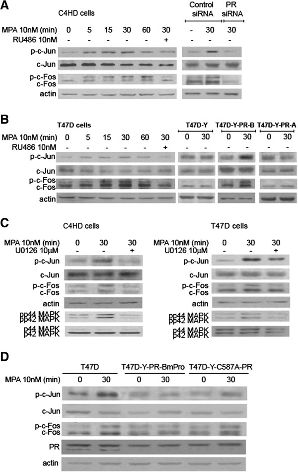
Results: We demonstrated that at the cyclin D1 promoter and through coordinated rapid and transcriptional effects, progestin induces the assembly of a transcriptional complex among AP-1, Stat3, PR, and ErbB-2 which functions as an enhanceosome to drive breast cancer growth. Our studies in a cohort of human breast tumors identified PR and AP-1 nuclear interaction as a marker of good prognosis and better OS in patients treated with tamoxifen (Tam), an anti-estrogen receptor therapy. Rationale for this finding was provided by our demonstration that Tam inhibits rapid and genomic PR effects, rendering breast cancer cells sensitive to its antiproliferative effects.

Conclusions: We here provided novel insight into the paradox of PR action as well as new tools to identify the subgroup of ER+/PR + patients unlikely to respond to ER-targeted therapies.


Figures

Figure 1
Figure 1
MPA induces c-Jun and c-Fos phosphorylation and AP-1 activation via p42/p44 MAPKs. (A) to (D) Cells were pretreated with RU486 or U0126, transfected with PR siRNAs or PR expression vectors and were then treated with MPA. Western blots (WB) were performed with phospho (p)-c-Jun and pp42/44MAPKs antibodies and filters were re-probed with the respective total antibody, or with a c-Fos antibody and re-probed with an actin antibody. Experiments in A to D were repeated five times with similar results. Signal intensities of phospho-proteins were analyzed by densitometry and normalized to total protein bands. Data analysis showed a significant increase in protein phosphorylation by MPA in comparison with untreated cells and a significant inhibition of MPA-induced phosphorylation by RU486 or UO126 (P <0.001). See also Additional file 1: Figure 1. MAPKs, Mitogen-activated protein kinases; MPA, Medroxyprogesterone acetate; PR, Progesterone receptor.
Figure 2
Figure 2
MPA induces c-Jun, c-Fos and PR nuclear colocalization and physical association. (A) PR, c-Jun and c-Fos were localized by IF and confocal microscopy. Merged images show MPA-induced c-Jun/PR or c-Fos/PR nuclear colocalization, evidenced by the yellow foci. Boxed areas are shown in detail in the right insets. Nuclei were stained with DAPI (blue). (B) PR/c-Jun and PR/-c-Fos nuclear interactions were detected by in situ PLA. The detected dimers are shown by the fluorescent rolling circle products (red). Nuclei were stained with DAPI (blue). The experiments shown were repeated three times with similar results. See also Additional file 1: Figure 2. MPA, Medroxyprogesterone acetate; PLA, Proximity ligation assay; PR, Progesterone receptor.
Figure 3
Figure 3
MPA modulates cyclin D1 expression via AP-1. (A) Cells were transfected with a cyclin D1 promoter luciferase construct containing the −954 TRE and with a construct with a point mutation in the TRE (−963 mut AP-1). When indicated, cells were co-transfected with TAM-67 or A-Fos and were then treated with MPA or pretreated with RU or U0 before MPA stimulation. As control of PR transcriptional activity cells were transfected with a PRE-Luc plasmid and stimulated with MPA. Results are presented as fold induction of luciferase activity with respect to cells untreated with MPA. Data represent the mean of three independent experiments for each cell type ± SEM. For b vs. a, and c vs. b: P <0.001. pA3 Luc, empty vector. MPA induces cyclin D1 expression at protein and mRNA levels via AP-1. Cells were transfected with TAM-67 and A-Fos vectors (B) and with c-Jun and c-Fos siRNAs (C) and then treated with MPA. Cyclin D1 protein expression was analyzed by WB. (D) Cyclin D1 mRNA expression levels were determined by RT-qPCR. The fold change of mRNA levels upon MPA treatment was calculated by normalizing the absolute levels of cyclin D1 mRNA to GAPDH levels, which was used as internal control, and setting the value of untreated cells as 1. Experiments shown were repeated three times with similar results. MPA, Medroxyprogesterone acetate.
Figure 4
Figure 4
MPA induces in vivo binding of c-Jun, c-Fos and PR to the cyclin D1 promoter. (A) Protein recruitment to the cyclin D1 promoter was analyzed by ChIP in cells treated with MPA or pretreated with U0126 when indicated. Immunoprecipitated DNA was amplified by qPCR using primers flanking the TRE site. The arbitrary qPCR number obtained for each sample was normalized to the input, setting the value of the untreated sample as 1. Data are expressed as n-fold chromatin enrichment over untreated cells. For b vs. a and c vs. b: P <0.001. (B) Sequential ChIP chromatins from cells treated with MPA were first immunoprecipitated with c-Jun or c-Fos antibodies and were then re-immunoprecipitated using a PR antibody. qPCR and data analysis were performed as detailed in A. For b vs. a: P <0.001. Results in A and B are the mean ± SEM from three independent experiments. IgG was used as a negative control. MPA effects in T47D-Y-C587A-PR cells. (C) Protein recruitment to the cyclin D1 promoter was studied as described in A. For b vs. a: P <0.001. (D) Cyclin D1 promoter activation was detected as in Figure  3A and data shown represent the mean of three independent experiments ± SEM. (E) Cyclin D1 expression was studied by WB. This experiment was repeated three times with similar results. ChIP, chromatin immunoprecipitation; MPA, Medroxyprogesterone acetate; PR, Progesterone receptor; WB, Western blot.
Figure 5
Figure 5
MPA induces a cooperative transcriptional interaction at the cyclin D1 promoter among AP-1, Stat3, PR and ErbB-2. (A) Cells were transfected with the indicated siRNAs or expression vectors and were then treated with MPA for 30 minutes. Recruitment of proteins to the cyclin D1 promoter was analyzed by ChIP. Immunoprecipitated DNA was amplified by qPCR using primers (red arrows) flanking the GAS and TRE sites indicated in the top panels. Amounts of immunoprecipitated DNA were normalized to inputs and reported relative to the amount obtained by IgG immunoprecipitation, which was set to one. (B) Sequential ChIP. Chromatins from cells treated with MPA as described in A were first immunoprecipitated with c-Jun or c-Fos antibodies and were then re-immunoprecipitated using an ErbB-2 antibody. The arbitrary qPCR number obtained for each sample was normalized to the input, setting the value of the untreated sample as 1. Data are expressed as fold chromatin enrichment over untreated cells. For b vs. a: P <0.001. (C) Recruitment of CBP and p300, and H3 and H4 acetylation levels (AcH3 and AcH4) at the sites described in A were studied by ChIP and data were also analyzed as in A. Results in A to C are the mean ± SEM from three independent experiments. For b vs. a: P <0.001. ChIP, chromatin immunoprecipitation; MPA, Medroxyprogesterone acetate.
Figure 6
Figure 6
AP-1/Stat3/PR/ErbB-2 transcriptional complex drives progestin-induced breast cancer growth. (A) and (B) Cells transfected as indicated were treated for 48 (C4HD) or 24 (T47D) h with MPA. Incorporation of [3H]thymidine was measured. Data are presented as the mean ± SD, P <0.001 for b vs. a and c vs. b. Experiments shown are representative of three. (C) Cyclin D1 protein expression in C4HD cells was analyzed by WB. (D) AP-1 activity and ErbB-2 nuclear function cooperate to drive in vivo progestin-induced growth. Left, cells (106) from each group were inoculated s.c. in mice treated with MPA and tumor volume was calculated as described in Methods. Each point represents tumor mean volume ± SEM. Right, decrease in tumor mass. (E) Tumor growth. aGrowth rates were calculated as the slopes of growth curves. Volume, percentage of growth inhibition and growth delay in tumors from the experimental groups with respect to tumors from control C4HD-p-Flag cells were calculated at Day 27. # vs. * and c vs. b for tumor volume and growth rate, P <0.001. d With respect to C4HD-p-Flag cells and f vs. e, P <0.001. g With respect to C4HD-p-Flag cells, P <0.001. (F) ChIP analysis. DNA-protein complexes were pulled down with the c-Jun antibody or with IgG and DNA was amplified by qPCR using primers indicated in Figure 5. Results are expressed as in Figure 5A and represent the average of three replicates ± SEM. For b vs. a, P <0.001. Shown is a representative sample of each tumor type. (G) Tumor lysates were analyzed by WB. C4HD cells growing in absence of MPA are shown as control: Shown are two representative samples of mice injected with the different experimental groups. See also Additional file 1: Figure 3. ChIP, chromatin immunoprecipitation; MPA, Medroxyprogesterone acetate; WB, Western blot.
Figure 7
Figure 7
PR and AP-1 interaction clinical significance. PR and p-c-Jun colocalization in tumor samples. (A) Nuclear p-c-Jun and PR levels were evaluated by IF and scored as described in Results. Protein colocalization was visualized as nuclear yellow dots, indicated by white arrows. Shown are examples of tumors showing 0 to +3 colocalization scores. Nuclei were stained with DAPI (blue). (B) Kaplan-Meier survival analysis correlating levels of p-c-Jun and PR colocalization with overall patient survival. IF, Immunofluorescence; PR, progesterone receptor.
Figure 8
Figure 8
PR and AP-1 interaction involvement in endocrine therapy response. (A) Proliferation , c-Jun phosphorylation and cyclin D1 promoter activation were studied as described in Figures 1, 3 and 6. (B) c-Jun and PR recruitment to the cyclin D1promoter was analyzed by ChIP as in Figure 5. Data are expressed as n-fold chromatin enrichment over untreated cells. For b vs. a and c vs. b: P <0.001. (C) to (J) Tam effects in sensitive and resistant cells. (C) and (D) Cell variants were treated as shown and proliferation was studied as in Figure 6. (E) Protein levels were analyzed by WB. Signal intensities of PR-A and PR-B bands were analyzed by densitometry and normalized to β-tubulin. Densitometric analysis of PR-A and PR-B expression levels in HR and HR6 clones, relative to those in BT474 cells (set to 1), are shown in the right panel. (F) and (G) WB in BT474-HR (F) and BT474 cells (G) were performed with the indicated phospho-antibodies and filters were re-probed with the respective total antibody. Signal intensities of phospho-proteins were normalized to total protein bands. Significance of MPA and Tam effects on the regulation of protein phosphorylation was analyzed as described in Methods (P <0.001). (H) and (I) c-Jun, PR, and ER α recruitment to the cyclin D1 promoter was studied by ChIP. We set as 1 the value of the untreated sample for BT474-HR cells (H) and of the IgG for BT474 (I). For b vs. a and c vs. b: P <0.001. (J) Tam effects on cyclin D1 protein expression. WBs were performed as in Figure 3 using β tubulin as loading control. Experiments in A to J were repeated five times with similar results. See Additional file 1: Figures 4, 5 and 6. ChIP, chromatin immunoprecipitation; MPA, Medroxyprogesterone acetate; PR, Progesterone receptor; Tam, Tamoxifen WB, Western blot.
Figure 9
Figure 9
PR and p-c-Jun colocalization in BT474 cell variants. Nuclear p-c-Jun and PR levels were evaluated by IF and scored as described in Results and shown in Figure 7A. Protein colocalization was visualized as nuclear yellow dots. Nuclei were stained with DAPI (blue). IF, Immunofluorescence; PR, Progesterone receptor.
Figure 10
Figure 10
Model of coordinated rapid and transcriptional PR effects that leads to the assembly of the AP-1/Stat3/ErbB-2/PR enhanceosome governing cyclin D1 expression. PR, Progesterone receptor.

References

    1. Daniel AR, Hagan CR, Lange CA. Progesterone receptor action: defining a role in breast cancer. Expert Rev Endocrinol Metab. 2011;15:359–369. doi: 10.1586/eem.11.25. - DOI - PMC - PubMed
    1. Balana ME, Lupu R, Labriola L, Charreau EH, Elizalde PV. Interactions between progestins and heregulin (HRG) signaling pathways: HRG acts as mediator of progestins proliferative effects in mouse mammary adenocarcinomas. Oncogene. 1999;15:6370–6379. doi: 10.1038/sj.onc.1203028. - DOI - PubMed
    1. Migliaccio A, Piccolo D, Castoria G, Di Domenico M, Bilancio A, Lombardi M, Gong W, Beato M, Auricchio F. Activation of the Src/p21ras/Erk pathway by progesterone receptor via cross-talk with estrogen receptor. EMBO J. 1998;15:2008–2018. doi: 10.1093/emboj/17.7.2008. - DOI - PMC - PubMed
    1. Boonyaratanakornkit V, Scott MP, Ribon V, Sherman L, Anderson SM, Maller JL, Miller WT, Edwards DP. Progesterone receptor contains a proline-rich motif that directly interacts with SH3 domains and activates c-Src family tyrosine kinases. Mol Cell. 2001;15:269–280. doi: 10.1016/S1097-2765(01)00304-5. - DOI - PubMed
    1. Carnevale RP, Proietti CJ, Salatino M, Urtreger A, Peluffo G, Edwards DP, Boonyaratanakornkit V, Charreau EH, Bal de Kier JE, Schillaci R, Elizalde PV. Progestin effects on breast cancer cell proliferation, proteases activation, and in vivo development of metastatic phenotype all depend on progesterone receptor capacity to activate cytoplasmic signaling pathways. Mol Endocrinol. 2007;15:1335–1358. doi: 10.1210/me.2006-0304. - DOI - PubMed

Publication types

MeSH terms