. 2014 Mar 1;55(2):218-27.
doi: 10.1093/jrr/rrt107. Epub 2013 Dec 17.

Static magnetic fields modulate X-ray-induced DNA damage in human glioblastoma primary cells

Affiliations

Static magnetic fields modulate X-ray-induced DNA damage in human glioblastoma primary cells

Laura Teodori et al. J Radiat Res. .

Abstract

Although static magnetic fields (SMFs) are used extensively in the occupational and medical fields, few comprehensive studies have investigated their possible genotoxic effect and the findings are controversial. With the advent of magnetic resonance imaging-guided radiation therapy, the potential effects of SMFs on ionizing radiation (IR) have become increasingly important. In this study we focused on the genotoxic effect of 80 mT SMFs, both alone and in combination with (i.e. preceding or following) X-ray (XR) irradiation, on primary glioblastoma cells in culture. The cells were exposed to: (i) SMFs alone; (ii) XRs alone; (iii) XR, with SMFs applied during recovery; (iv) SMFs both before and after XR irradiation. XR-induced DNA damage was analyzed by Single Cell Gel Electrophoresis assay (comet assay) using statistical tools designed to assess the tail DNA (TD) and tail length (TL) as indicators of DNA fragmentation. Mitochondrial membrane potential, known to be affected by IR, was assessed using the JC-1 mitochondrial probe. Our results showed that exposure of cells to 5 Gy of XR irradiation alone led to extensive DNA damage, which was significantly reduced by post-irradiation exposure to SMFs. The XR-induced loss of mitochondrial membrane potential was to a large extent averted by exposure to SMFs. These data suggest that SMFs modulate DNA damage and/or damage repair, possibly through a mechanism that affects mitochondria.

Keywords: DNA fragmentation; comet assay; glioblastoma; ionizing radiation; mitochondrial membrane potential; static magnetic field.


Figures

Fig. 1.
Fig. 1.
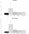
Comet assay results of cells exposed to SMFs. The percentage of DNA in tail (TD) and 90% of tail length (TL) are shown in the upper and lower panel, respectively. The results of triplicate samples of two independent experiments are reported together with the SD for each time-point. Cells were exposed to an 80 ± 5 mT SMF in exponential growth for 6, 12 and 24 h. A statistically significant difference was observed (asterisk) between sham- and SMF-exposed cells after 24 h, and within both sham and exposed groups between 6 and 24 h of treatment (both TD and TL parameters) (P < 0.01).
Fig. 2.
Fig. 2.
Comet assay results of cells exposed to XR irradiation that underwent 6 h of recovery in the absence and in the presence of SMFs. TD and TL are shown in the upper and lower panels, respectively. The results of triplicate samples of two independent experiments are reported together with the SD. Five groups are reported in the figure. These groups are (i) sham: cells not exposed to any treatment but submitted to the same environmental conditions; (ii) XR: cells exposed to XR irradiation only; (iii) SMF: cells exposed to SMFs only; (iv) XR + SMF: cells exposed to SMFs during 6 h of recovery following XR irradiation; (v) SMF + XR + SMF: cells exposed to SMFs prior to XR irradiation and during 6 h of recovery. Recovery under SMFs significantly (asterisk) reduced the TD and TL (P < 0.001) compared with XR irradiation alone.
Fig. 3.
Fig. 3.
Comet test results for cells exposed to 5 Gy XR irradiation that subsequently underwent 20 h of recovery with or without SMFs. TD and TL are shown in the upper and lower panels, respectively. The results of triplicate samples of two independent experiments are reported together with the SD. Five groups are reported in the figure. These groups are (i) sham: cells not exposed to any treatment but submitted to the same environmental conditions; (ii) XR: cells exposed to XR irradiation only; (iii) SMF: cells exposed to SMF only; (iv) XR + SMF: cells exposed to SMF during 20 h of recovery following XR irradiation; (v) SMF + XR + SMF: cells exposed to SMFs prior to XR irradiation and during 20 h of recovery. Recovery under SMFs significantly (asterisk) reduced the MDL and TL (P < 0.001) compared with XR irradiation alone.
Fig. 4.
Fig. 4.
Representative histogram of JC-1 590 nm fluorescence, indicative of the ΔΨm induced by the treatment, analyzed after 6 h of recovery following XR irradiation. Each fluorescence group is highlighted with a different color. A shift toward a reduced 590 fluorescence is evident in the XR-irradiated cells (green curve) compared with the sham cells (yellow curve). The presence of SMFs during the recovery phase following XR irradiation, instead, reduced the ΔΨm (blue curve).
Fig. 5.
Fig. 5.
Time-course of ΔΨm variation as determined by JC-1 staining. The figure shows the ratios between the positive and negative 590 nm fluorescent cells measured after 3, 6 and 20 h of recovery following XR irradiation, in the presence of SMFs (XR + SMF) or absence of SMFs (XR). The above ratios of cells exposed to SMFs alone (SMF) and of sham cells (SHAM) are also reported.

References

    1. Schiffer IB, Schreiber WG, Graf R, et al. No influence of magnetic fields on cell cycle progression using conditions relevant for patients during MRI. Bioelectromagnetics. 2003;24:241–50. - PubMed
    1. Teodori L, Albertini MC, Uguccioni F, et al. Static magnetic fields affect cell size, shape, orientation, and membrane surface of human glioblastoma cells, as demonstrated by electron, optic, and atomic force microscopy. Cytometry A. 2006;69:75–85. - PubMed
    1. Coletti D, Teodori L, Albertini MC, et al. Static magnetic fields enhance skeletal muscle differentiation in vitro by improving myoblast alignment. Cytometry A. 2007;71:846–56. - PubMed
    1. Tenuzzo B, Chionna A, Panzarini E, et al. Biological effects of 6 mT static magnetic fields: a comparative study in different cell types. Bioelectromagnetics. 2006;27:560–77. - PubMed
    1. Chekhun VF, Lozovskaya YV, Lukyanova NY, et al. Assessment of the cyto- and genotoxic effects of a nanoferromagnetic and a static magnetic field in vivo. Cytol Genet. 2013;47:179–87.

Publication types