. 2013 Dec 17;6(306):ra108.
doi: 10.1126/scisignal.2004405.

PLCε, PKD1, and SSH1L transduce RhoA signaling to protect mitochondria from oxidative stress in the heart

Affiliations

PLCε, PKD1, and SSH1L transduce RhoA signaling to protect mitochondria from oxidative stress in the heart

Sunny Y Xiang et al. Sci Signal. .

Abstract

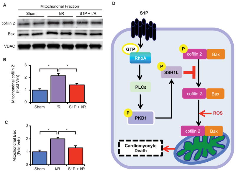
Activation of the small guanosine triphosphatase RhoA can promote cell survival in cultured cardiomyocytes and in the heart. We showed that the circulating lysophospholipid sphingosine 1-phosphate (S1P), a G protein (heterotrimeric guanine nucleotide-binding protein)-coupled receptor (GPCR) agonist, signaled through RhoA and phospholipase Cε (PLCε) to increase the phosphorylation and activation of protein kinase D1 (PKD1). Genetic deletion of either PKD1 or its upstream regulator PLCε inhibited S1P-mediated cardioprotection against ischemia/reperfusion injury. Cardioprotection involved PKD1-mediated phosphorylation and inhibition of the cofilin phosphatase Slingshot 1L (SSH1L). Cofilin 2 translocates to mitochondria in response to oxidative stress or ischemia/reperfusion injury, and both S1P pretreatment and SSH1L knockdown attenuated translocation of cofilin 2 to mitochondria. Cofilin 2 associates with the proapoptotic protein Bax, and the mitochondrial translocation of Bax in response to oxidative stress was also attenuated by S1P treatment in isolated hearts or by knockdown of SSH1L or cofilin 2 in cardiomyocytes. Furthermore, SSH1L knockdown, like S1P treatment, increased cardiomyocyte survival and preserved mitochondrial integrity after oxidative stress. These findings reveal a pathway initiated by GPCR agonist-induced RhoA activation, in which PLCε signals to PKD1-mediated phosphorylation of cytoskeletal proteins to prevent the mitochondrial translocation and proapoptotic function of cofilin 2 and Bax and thereby promote cell survival.


Conflict of interest statement

Competing interests: The authors declare that they have no competing interests.

Figures

Fig. 1
Fig. 1
S1P activates RhoA and PKD1, and PKD1 gene deletion prevents S1P protection in the heart. (A and B) Mouse hearts were perfused with S1P or Vehicle (Veh) and RhoA activation and PKD1 phosphorylation in the left ventricle were determined. (A) Quantification of GTP RhoA amount. n = 4 animals per group. (B) Representative blots (top) and quantification (bottom) of phosphorylation of PKD1 (Ser744/748). n = 5 animals per group. (C) Representative blots showing PKD1 protein abundance in WT and PKD1 KO mouse hearts. (D) WT and PKD1 KO mouse hearts were subjected to ischemia/reperfusion injury with Veh or S1P pretreatment. Representative images of TTC stained cross sections of heart following ischemia/reperfusion injury (top) and quantification of the infarct size (bottom). n = 5–6 animals per group.
Fig. 2
Fig. 2
S1P activates PKD1 through RhoA and PLCε, and PLCε gene deletion prevents S1P protection in the heart. (A and B) Time course phosphorylation of PKD1 in response to S1P and inhibition by C3 exoenzyme (A) and PLCε knockdown with siRNA (B) in cardiomyocytes. n = 3–5 experiments per time point. (C) WT and PLCε KO mouse hearts were perfused with Veh or S1P for 30 min. Representative blots (top) and quantification (bottom) showing PKD1 phosphorylation. n = 5 animals per group. (D) WT and PLCε KO mouse hearts were subjected to ischemia/reperfusion injury with Veh or S1P pretreatment. Representative images of TTC stained cross sections of heart following ischemia/reperfusion injury (top) and quantification of the infarct size (bottom). n = 5–6 animals per group.
Fig. 3
Fig. 3
PKD1 dependent phosphorylation of SSH1L and cofilin 2 in cardiomyocytes. (A and B) Representative blots (top) and quantification (bottom) of dose-dependent phosphorylation of SSH1L (A) and cofilin 2 (B) by 30 min S1P treatment. n = 4 experiments per time point. (C) Cardiomyocytes were transfected with Ctr siRNA or SSH1L siRNA. Representative blots showing cofilin abundance, phosphorylation, and SSH1L abundance (top) and quantification of cofilin 2 phosphorylation after SSH1L knockdown (bottom). n = 4 experiments per treatment. (D and E) Cardiomyocytes were transfected with Ctr siRNA or PKD1 siRNA then subjected to S1P treatment. Time course of the phosphorylation of SSH1L (D) and cofilin 2 (E) by S1P. n = 4–5 experiments per time point. (F) Hypothetical scheme for S1P-PKD1-SSH1L-cofilin2 signaling.
Fig. 4
Fig. 4
PKD1 mediates S1P induced cell survival and SSH1L knockdown mimics S1P protection in response to H2O2 treatment in cardiomyocytes. (A through C) Cardiomyocytes were transfected with Ctr siRNA or PKD1 siRNA. (A) Representative images of cardiomyocytes stained with Calcein (green) and propidium iodide (red) following indicated treatments. Scale bar, 40 μm. Quantification of cardiomyocyte viability (B) and LDH release (C) following the indicated treatments. (D and E) Cardiomyocytes were transfected with Ctr siRNA or SSH1L siRNA. Quantification of cardiomyocyte viability (D) and LDH release (E) following the indicated treatments. n = 6 experiments per treatment for (B) to (E).
Fig. 5
Fig. 5
S1P or SSH1L knockdown decreases cytochrome c release and preserves mitochondrial membrane potential in response to H2O2 treatment in cardiomyocytes. (A and B) Cytochrome c (Cyto c) release in the cytosolic fraction with H2O2. (A) Representative blots (top) and quantification (bottom) of Cyto c release with or without S1P pretreatment. (B) Representative blots (top) and quantification (bottom) of Cyto c release in Ctr siRNA or SSH1L siRNA transfected cells. (C and D) TMRE fluorescence intensity in response to H2O2 treatment. (C) Representative images of TMRE staining (top) and quantification (bottom) of TMRE fluorescence intensity with or without S1P pretreatment. Scale bar, 20 μm. (D) Representative images of TMRE staining (top) and quantification (bottom) of TMRE fluorescence intensity in Ctr siRNA or SSH1L siRNA transfected cells. Scale bar, 20 μm. n = 6 experiments per treatment for (A) to (D).
Fig. 6
Fig. 6
S1P and SSH1L regulate mitochondrial translocation of cofilin 2 and Bax in H2O2-treated cardiomyocytes. (A) Cardiomyocytes were pretreated with S1P or Veh and then subjected to H2O2 treatment. Representative blots (top) and quantification (bottom) of mitochondrial abundance of cofilin 2. (B) Cardiomyocytes were transfected with Ctr siRNA or SSH1L siRNA then subjected to H2O2 treatment. Representative blots (top) and quantification (bottom) of mitochondrial abundance of cofilin 2. (C) Representative blots showing co-immunoprecipitation of cofilin 2 and Bax. (D) Cardiomyocytes were pretreated with S1P or Veh and then subjected to H2O2 treatment. Representative blots (top) and quantification (bottom) of mitochondrial Bax. (E) Cardiomyocytes were transfected with Ctr siRNA or SSH1L siRNA then subjected to H2O2 treatment. Representative blots (top) and quantification (bottom) of mitochondrial Bax. (F) Cardiomyocytes were transfected with Ctr siRNA or cofilin 2 siRNA then subjected to H2O2 treatment. Representative blots (top) and quantification (bottom) of mitochondrial Bax. n = 5–6 experiments per treatment for (A) to (F).
Fig. 7
Fig. 7
S1P attenuates mitochondrial translocation of cofilin 2 and Bax by ischemia/reperfusion injury in the isolated heart. Isolated mouse hearts were perfused with Veh or S1P prior to 20 min ischemia followed by 30 min reperfusion. (A) Representative blots showing mitochondrial cofilin 2 and Bax abundance. (B) Quantification of mitochondrial cofilin 2 and (C) mitochondrial Bax. n = 4–6 animals per group. (D) Hypothetical schema showing the role of the S1P-RhoA-PKD1-SSH1L signaling pathway in oxidative stress induced cofilin 2 and Bax translocation to mitochondria and cardiomyocyte cell death.

References

    1. Hart MJ, Jiang X, Kozasa T, Roscoe W, Singer WD, Gilman AG, Sternweis PC, Bollag G. Direct stimulation of the guanine nucleotide exchange activity of p115 RhoGEF by Galpha13. Science. 1998 Jun 26;280:2112–2114. - PubMed
    1. Kozasa T, Jiang X, Hart MJ, Sternweis PM, Singer WD, Gilman AG, Bollag G, Sternweis PC. p115 RhoGEF, a GTPase activating protein for Galpha12 and Galpha13. Science. 1998 Jun 26;280:2109–2111. - PubMed
    1. Siehler S, Manning DR. Pathways of transduction engaged by sphingosine 1-phosphate through G protein-coupled receptors. Biochim Biophys Acta. 2002 May 23;1582:94–99. - PubMed
    1. Sugimoto N, Takuwa N, Okamoto H, Sakurada S, Takuwa Y. Inhibitory and stimulatory regulation of Rac and cell motility by the G12/13-Rho and Gi pathways integrated downstream of a single G protein-coupled sphingosine-1-phosphate receptor isoform. Mol Cell Biol. 2003 Mar;23:1534–1545. - PMC - PubMed
    1. Xiang SY, Dusaban SS, Brown JH. Lysophospholipid receptor activation of RhoA and lipid signaling pathways. Biochim Biophys Acta. 2013 Jan;1831:213–222. - PMC - PubMed

Publication types

MeSH terms