. 2013 Dec 17;2(12):e139.
doi: 10.1038/mtna.2013.66.

Influence of Polyethylene Glycol Lipid Desorption Rates on Pharmacokinetics and Pharmacodynamics of siRNA Lipid Nanoparticles

Affiliations

Influence of Polyethylene Glycol Lipid Desorption Rates on Pharmacokinetics and Pharmacodynamics of siRNA Lipid Nanoparticles

Barbara L Mui et al. Mol Ther Nucleic Acids. .

Abstract

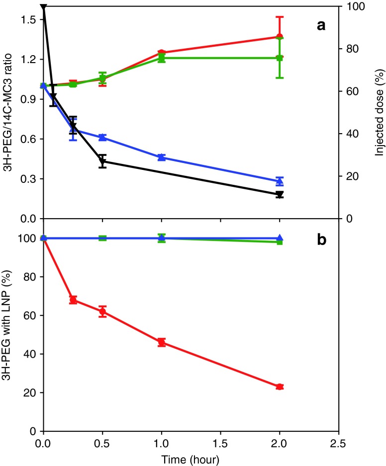
Lipid nanoparticles (LNPs) encapsulating short interfering RNAs that target hepatic genes are advancing through clinical trials, and early results indicate the excellent gene silencing observed in rodents and nonhuman primates also translates to humans. This success has motivated research to identify ways to further advance this delivery platform. Here, we characterize the polyethylene glycol lipid (PEG-lipid) components, which are required to control the self-assembly process during formation of lipid particles, but can negatively affect delivery to hepatocytes and hepatic gene silencing in vivo. The rate of transfer from LNPs to plasma lipoproteins in vivo is measured for three PEG-lipids with dialkyl chains 14, 16, and 18 carbons long. We show that 1.5 mol % PEG-lipid represents a threshold concentration at which the chain length exerts a minimal effect on hepatic gene silencing but can still modify LNPs pharmacokinetics and biodistribution. Increasing the concentration to 2.5 and 3.5 mol % substantially compromises hepatocyte gene knockdown for PEG-lipids with distearyl (C18) chains but has little impact for shorter dimyristyl (C14) chains. These data are discussed with respect to RNA delivery and the different rates at which the steric barrier disassociates from LNPs in vivo.Molecular Therapy-Nucleic Acids (2013) 2, e139; doi:10.1038/mtna.2013.66; published online 17 December 2013.


Figures

Figure 1
Figure 1
Plasma and tissue concentration-time profiles at different dose levels. LNPs containing 1.5 mol % PEG-C14 and trace 3H-CHE were administered to mice at doses of 0.33 (circle), 3.3 (square), and 11.1 (triangle) mg/kg of lipid, equivalent to 0.03, 0.3, and 1 mg/kg siRNA, respectively. At various times, the animals were sacrificed and the amount of radiolabel present in blood, liver, and spleen was determined as outlined in Materials and Methods section. Dose proportionality is highlighted by the inserted linear plots of AUC (ng lipid·hour/g) versus lipid dose (mg/kg). Data points represent the average of 4 animals ± 1 SD. AUC, area under the curve; LNP, lipid nanoparticles; PEG, polyethylene glycol.
Figure 2
Figure 2
Comparison of 14C-MC3 and 3H-CHE as LNP markers. Mice, injected with LNPs containing 1.5 mol % PEG-C14, dual labeled with 14C-MC3 and 3H-CHE, were sacrificed at various times up to a maximum of 2 hours and the amount of radiolabel present in the blood determined as outlined in Materials and Methods section. The % injected dose was determined using 3H-CHE (square) and the 14C-MC3 to 3H-CHE ratio (circle) was used to show 14C-MC3 is a stable LNP marker for at least 2 hours after injection, during which time ~90% of the injected dose distributes out of the blood compartment. Data represent the average of 4 animals ± 1 SD. LNP, lipid nanoparticles; PEG, polyethylene glycol.
Figure 3
Figure 3
Loss of PEG-lipid from LNPs in circulation. (a) Mice injected with LNPs containing 1.5% mol % PEG-C14, dual labeled with 14C-MC3 and 3H-PEG-C14, were sacrificed at various times and the 3H-PEG to 14C-MC3 ratio determined in blood (circle), plasma (square), and LNP isolated from plasma (triangle) as outlined in Materials and Methods section. The clearance of the LNP as measured by 14C-MC3 in the blood is shown as the percent injected dose (inverted triangle). (b) LNPs containing 1.5 mol % PEG-C14 (circle), PEG-C16 (square), or PEG-C18 (triangle), dual labeled with 14C-MC3 and the corresponding 3H-PEG-lipid were injected into mice. At various times the animals were sacrificed, plasma isolated and the amount of 3H-PEG retained with LNPs measured as outlined in Materials and Methods section. Data represent the average of 4 animals ± 1 SD. LNP, lipid nanoparticles; PEG, polyethylene glycol.
Figure 4
Figure 4
Effect of PEG-lipid on FVII gene silencing activity. (a) FVII siRNA LNPs containing 1.5 mol % PEG-C14 (circle), PEG-C16 (triangle), or PEG-C18 (square) were administered to mice at the indicated siRNA doses and plasma FVII protein concentrations determined 24 hours later. ED50's of 0.02, 0.03, and 0.04 mg/kg siRNA estimated for PEG-C14, PEG-C16, and PEG-C18 respectively are not significantly different (P > 0.05). (b) PEG-C14 content of 1.5 mol % (circle), 2.5% (triangle), and 3.5% (square) exhibit ED50's of 0.02, 0.03, and 0.06 mg/kg siRNA respectively, which are also not significantly different. (c) PEG-C18 content of 1.5 mol % (circle), 2.5% (triangle), and 3.5% (square) exhibit ED50's of 0.04, >0.3, and >0.3 mg/kg siRNA, respectively. The difference in activity observed at 1.5% PEG-C18 is statistically significant compared to 2.5 and 3.5% (**P < 0.005). Data points represent the average of 3–6 mice ± 1 SD. LNP, lipid nanoparticles; PEG, polyethylene glycol.
Figure 5
Figure 5
Effect of PEG-lipid chain length on pharmacokinetics and biodistribution. Mice were administered LNPs containing 1.5 mol % PEG-C14 (diamond), PEG-C16 (square), and PEG-C18 (triangle), dual labeled with 14C-MC3 and the corresponding 3H-PEG-lipids. At various times the animals were sacrificed and the concentration of 14C and 3H measured in blood, liver, and spleen. Data points represent the average of 4 mice ± 1 SD. LNP, lipid nanoparticles; PEG, polyethylene glycol.

References

    1. Coelho T, Adams D, Silva A, Lozeron P, Hawkins PN, Mant T, et al. Safety and efficacy of RNAi therapy for transthyretin amyloidosis. N Engl J Med. 2013;369:819–829. - PubMed
    1. Fitzgerald K, Frank-Kamenetsky M, Shulga-Morskaya S, Liebow A, Bettencourt BR, Sutherland JE, et al. Effect of an RNA interference drug on the synthesis of proprotein convertase subtilisin/kexin type 9 (PCSK9) and the concentration of serum LDL cholesterol in healthy volunteers: a randomised, single-blind, placebo-controlled, phase 1 trial. Lancet. 2013. - PMC - PubMed
    1. de Fougerolles A, Vornlocher HP, Maraganore J, Lieberman J. Interfering with disease: a progress report on siRNA-based therapeutics. Nat Rev Drug Discov. 2007;6:443–453. - PMC - PubMed
    1. Zimmermann TS, Lee AC, Akinc A, Bramlage B, Bumcrot D, Fedoruk MN, et al. RNAi-mediated gene silencing in non-human primates. Nature. 2006;441:111–114. - PubMed
    1. Jayaraman M, Ansell SM, Mui BL, Tam YK, Chen J, Du X, et al. Maximizing the potency of siRNA lipid nanoparticles for hepatic gene silencing in vivo. Angew Chem Int Ed Engl. 2012;51:8529–8533. - PMC - PubMed