. 2014 Jan;6(1):28-33.
doi: 10.1038/nchem.1795. Epub 2013 Nov 24.

Oligomerization transforms human APOBEC3G from an efficient enzyme to a slowly dissociating nucleic acid-binding protein

Affiliations

Oligomerization transforms human APOBEC3G from an efficient enzyme to a slowly dissociating nucleic acid-binding protein

Kathy R Chaurasiya et al. Nat Chem. 2014 Jan.

Abstract

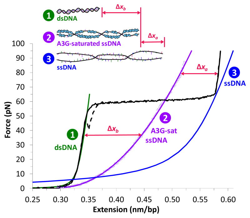
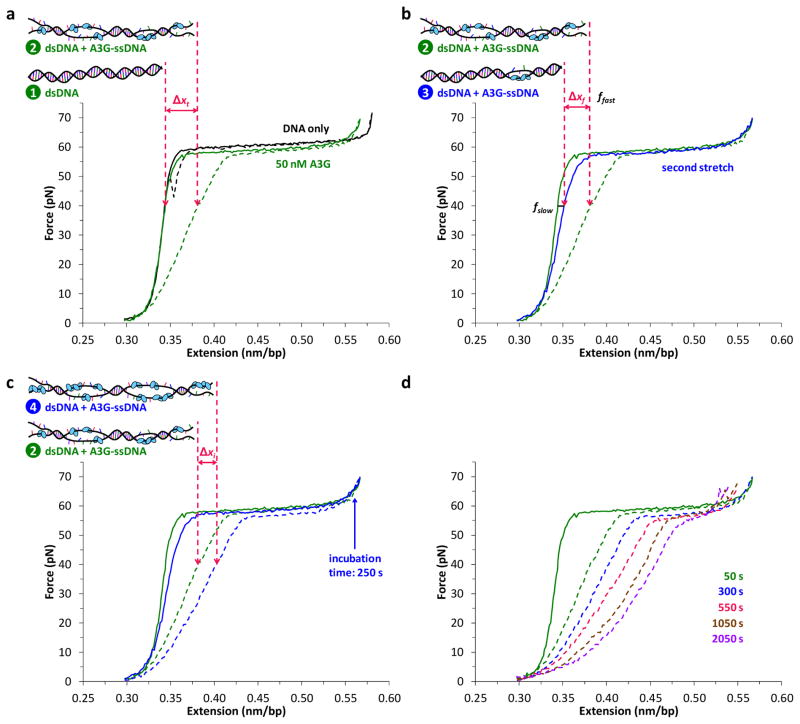
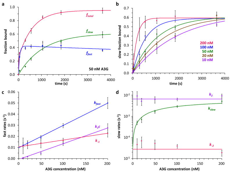
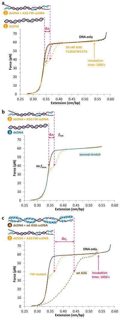
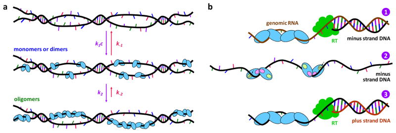
The human APOBEC3 proteins are a family of DNA-editing enzymes that play an important role in the innate immune response against retroviruses and retrotransposons. APOBEC3G is a member of this family that inhibits HIV-1 replication in the absence of the viral infectivity factor Vif. Inhibition of HIV replication occurs by both deamination of viral single-stranded DNA and a deamination-independent mechanism. Efficient deamination requires rapid binding to and dissociation from ssDNA. However, a relatively slow dissociation rate is required for the proposed deaminase-independent roadblock mechanism in which APOBEC3G binds the viral template strand and blocks reverse transcriptase-catalysed DNA elongation. Here, we show that APOBEC3G initially binds ssDNA with rapid on-off rates and subsequently converts to a slowly dissociating mode. In contrast, an oligomerization-deficient APOBEC3G mutant did not exhibit a slow off rate. We propose that catalytically active monomers or dimers slowly oligomerize on the viral genome and inhibit reverse transcription.


Figures

Figure 1
Figure 1
The force-dependent difference in length between DNA and a saturated A3G-DNA complex allows us to measure A3G binding. Typical extension (solid black) and return (dashed black) of a single DNA molecule. At 61.0 ± 0.5 pN, the molecule undergoes a force-induced melting transition from dsDNA (green, Eq. S1) to ssDNA (blue). A3G-saturated ssDNA (200 nM A3G, t > 500 s, data points fit to Eq. S4, solid purple) is longer than dsDNA (Δxb below the melting transition) and shorter than ssDNA (Δxa above the melting transition). A3G-saturated ssDNA is significantly shorter than ssDNA only (blue, Eq. S2), which suggests that A3G may wrap ssDNA upon binding.
Figure 2
Figure 2
Single molecule method to measure fast and slow fractions of A3G binding. (A) Without protein (black), a single DNA molecule reanneals immediately upon release, exhibiting minimal hysteresis, or mismatch between stretch (solid) and release (dashed) curves. In the presence of 50 nM A3G, the stretch curve (solid green) follows the dsDNA-only curve, indicating negligible A3G-dsDNA binding. A3G binds the exposed ssDNA, and prohibits the DNA strands from reannealing, resulting in hysteresis (dashed green). For a given force (40 pN shown), there is a corresponding change in DNA length Δxt between A3G-free dsDNA (left arrow, drawing 1) and partially A3G-bound ssDNA (right arrow, drawing 2). This force-dependent length change measures A3G-ssDNA binding (Supplementary Fig. 1). (B) The second stretch (solid blue) lies between the first stretch and release curves, distinguishing the fraction of A3G that remains bound (fslow) from the fraction that dissociated (ffast) before the second stretch. The A3G that dissociates rapidly allows the strands to reanneal immediately into dsDNA (drawing 3), resulting in length decrease Δxf. (C) Pausing at fixed DNA extension after incubating ssDNA with 50 nM A3G results in additional binding (drawing 4), indicated by the corresponding length increase Δxi measured during DNA release. (D) A3G binding increases with total exposure time to ssDNA (dashed lines).
Figure 3
Figure 3
Quantifying A3G binding reveals association and dissociation rates for fast and slow binding modes. (a) Total binding at 50 nM A3G (ftotal, red) separated into a fast fraction (ffast, blue) and slow fraction (fslow, green), as a function of ssDNA-A3G incubation time. Fits to the binding model (solid lines, Supplementary Eqs. S9–S11) yield observed rates kfast and kslow. (b) Slow fraction bound as a function of time for five A3G concentrations. Solid lines are fits to Supplementary Eq. S10. Error bars (panels a, b) are standard error (N≥3) for 50–200 nM A3G and propagated error for 10–20 nM A3G. (c) Fast rates (kfast, blue data points) obtained from fits to the binding model (shown in panel a for 50 nM A3G). The linear fit (solid blue line, Supplementary Eq. S13) yields k1 and k−1. k1c (purple data points) and k−1 (red data points) were also calculated from the binding model. Linear fits (solid lines, Supplementary Eqs. S15 and S17) yield consistent values of k1 and k−1. (d) Slow rates (kslow, green data points) from fits to the binding model (panel b). Fits to Supplementary Eq. S21 (solid green line, see Supplementary Fig. 2, panel b) yield k2 and k−2. Separate calculations of k2 (purple) and k−2 (red) from the binding model (Supplementary Eqs. S24 and S25) are also shown.
Figure 4
Figure 4
Oligomerization-defective mutant F126A/W127A (FW) demonstrates that the slow kinetics observed for wild type A3G is due to oligomerization. (a) In the absence of protein (black), a single DNA molecule reanneals immediately upon release, exhibiting minimal hysteresis between extension (solid) and release (dashed). In the presence of 50 nM F126A/W127A A3G (orange), the stretch curve (solid) follows the dsDNA-only curve, indicating no measurable A3G FW binding to dsDNA (drawing 1). Pausing at fixed DNA extension after the melting transition to incubate the ssDNA with the protein results in ssDNA binding (drawing 2), indicated by the corresponding increase in length Δxt measured during DNA release at a given force (shown for 40 pN). (b) The subsequent stretch (dark blue) follows the initial stretch curve (solid orange), indicating that all the mutant A3G bound during incubation dissociates rapidly (drawing 3), resulting in a decrease in length Δxf. (c) Wild type A3G (drawing 4) exhibits a greater change in length Δxo relative to the FW mutant (drawing 2) at 1050 s incubation due to oligomerization on ssDNA.
Figure 5
Figure 5
Models for A3G oligomerization (A) in vitro and (B) in virio. (a) Initially monomers or dimers bind ssDNA with on rate k1c and off rate k−1. These forward and backward rates are on similar timescales (1/k1c = 33 ± 1 s at 200 nM A3G, and 1/k−1 = 85 ± 5 s), so fast binding reaches equilibrium before the monomers or dimers convert to oligomers (1/k2 = 149 ± 13 s) on ssDNA. Oligomer dissociation is significantly slower (1/k−2 = 10 ± 2 h) in vitro. (b) Inside the virus, A3G oligomerizes on the RNA genome, blocking minus-strand DNA synthesis by RT (panel 1). Once the oligomer dissociates, the monomers or dimers released bind ssDNA within a second, allowing rapid enzymatic activity (panel 2) until A3G oligomerizes on the ssDNA template in 150 s and blocks plus strand synthesis (panel 3).

Comment in

References

    1. Malim MH. APOBEC proteins and intrinsic resistance to HIV-1 infection. Philos Trans R Soc Lond B Biol Sci. 2009;364(1517):675–687. - PMC - PubMed
    1. Harris RS, Liddament MT. Retroviral restriction by APOBEC proteins. Nat Rev Immunol. 2004;4(11):868–877. - PubMed
    1. Duggal NK, Emerman M. Evolutionary conflicts between viruses and restriction factors shape immunity. Nat Rev Immunol. 2012;12(10):687–695. - PMC - PubMed
    1. Chiu YL, Greene WC. The APOBEC3 cytidine deaminases: An innate defensive network opposing exogenous retroviruses and endogenous retroelements. Annu Rev Immunol. 2008;26:317–353. - PubMed
    1. Sheehy AM, Gaddis NC, Choi JD, Malim MH. Isolation of a human gene that inhibits HIV-1 infection and is suppressed by the viral Vif protein. Nature. 2002;418(6898):646–650. - PubMed

Publication types