. 2013 Dec 17;3(12):e338.
doi: 10.1038/tp.2013.105.

dcc orchestrates the development of the prefrontal cortex during adolescence and is altered in psychiatric patients

Affiliations

dcc orchestrates the development of the prefrontal cortex during adolescence and is altered in psychiatric patients

C Manitt et al. Transl Psychiatry. .

Abstract

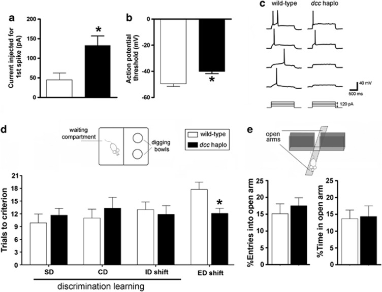
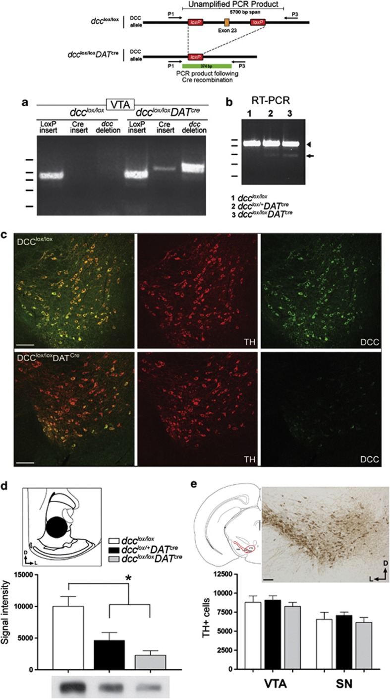
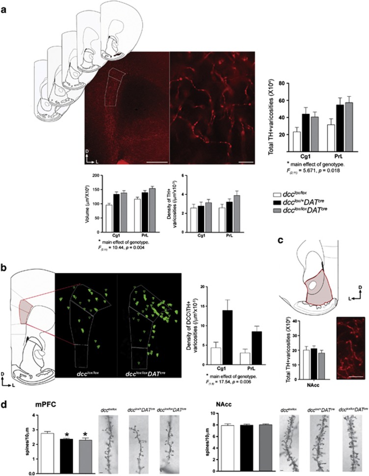
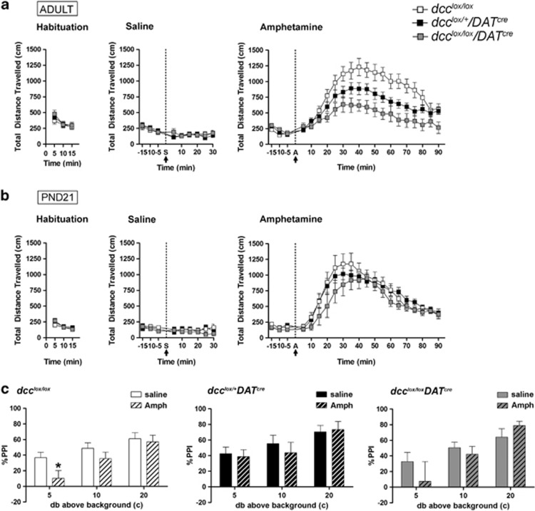
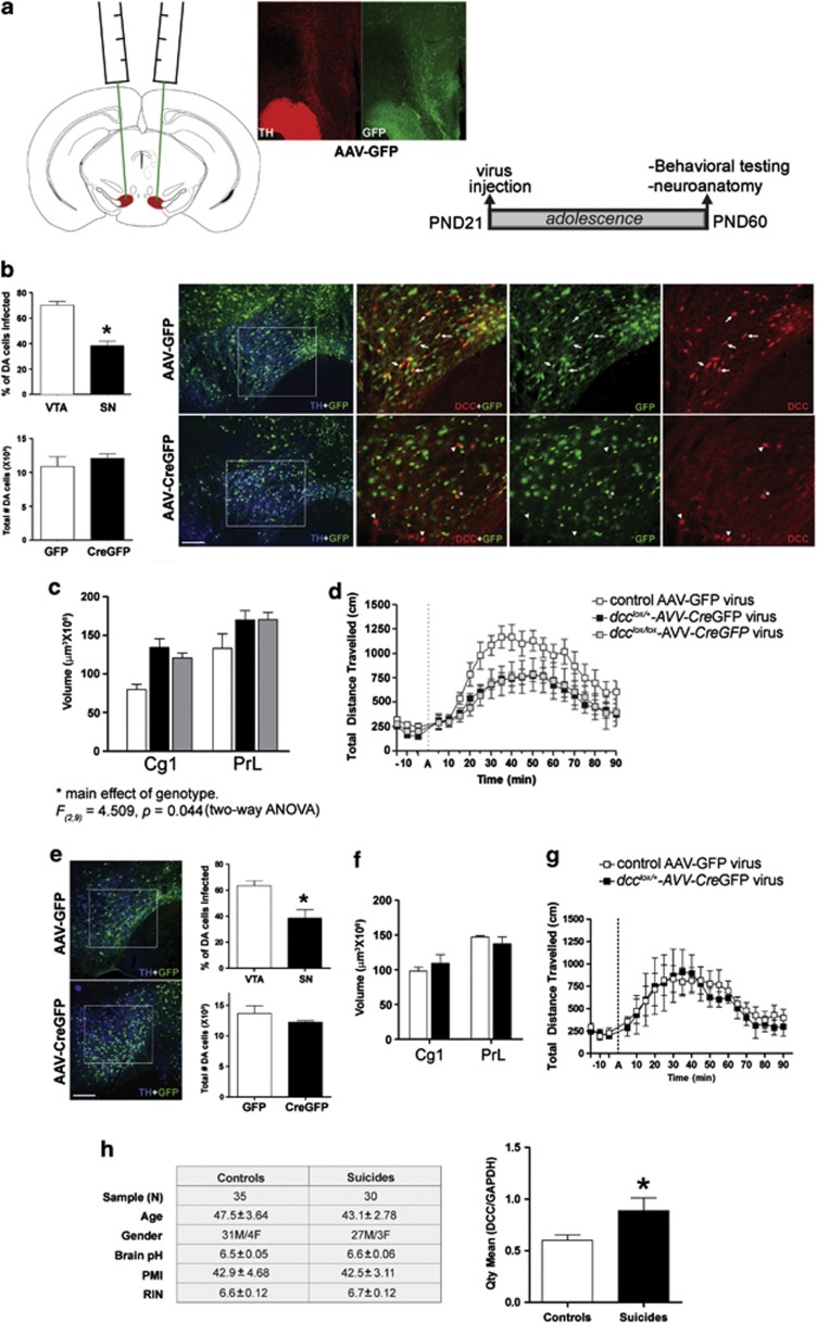
Adolescence is a period of heightened susceptibility to psychiatric disorders of medial prefrontal cortex (mPFC) dysfunction and cognitive impairment. mPFC dopamine (DA) projections reach maturity only in early adulthood, when their control over cognition becomes fully functional. The mechanisms governing this protracted and unique development are unknown. Here we identify dcc as the first DA neuron gene to regulate mPFC connectivity during adolescence and dissect the mechanisms involved. Reduction or loss of dcc from DA neurons by Cre-lox recombination increased mPFC DA innervation. Underlying this was the presence of ectopic DA fibers that normally innervate non-cortical targets. Altered DA input changed the anatomy and electrophysiology of mPFC circuits, leading to enhanced cognitive flexibility. All phenotypes only emerged in adulthood. Using viral Cre, we demonstrated that dcc organizes mPFC wiring specifically during adolescence. Variations in DCC may determine differential predisposition to mPFC disorders in humans. Indeed, DCC expression is elevated in brains of antidepressant-free subjects who committed suicide.


Figures

Figure 1
Figure 1
Improved behavioral flexibility and reduced excitability of layer V mPFC pyramidal neurons in dcc haploinsufficient mice. (ac) Depolarizing current steps (30pA) were injected into layer V pyramidal neurons of the pregenual mPFC (spanning the cingulate 1 and prelimbic subregions) to estimate the rheobase current, which was defined as the first current step capable of eliciting one action potential. (a) The average injected current that was required to trigger an action potential (1st spike) was significantly higher in adult dcc haploinsufficient mice compared with wild-type littermate controls (wild-type: n=6; dcc haploinsufficient: n=8; t(12)=3.013, P=0.0108). (b) The average action potential threshold of Layer V pyramidal neurons is significantly higher in dcc haploinsufficient mice (wild-type: n=6; dcc haploinsufficient: n=8; t(12)=3.788, P=0.0026. (c) Representative membrane potential traces recorded from mPFC layer V pyramidal neurons of wild-type and dcc haploinsufficient mice. Note that more current steps are needed to trigger action potential formation in dcc haplosinsufficient mice. (d) dcc haploinsufficient mice exhibit increased behavioral flexibility in the Attentional Set-Shifting Task. No differences were observed between dcc haploinsufficient and wild-type mice in the mean number of trials to criterion required to solve the SD, CD, and ID tasks, indicating no differences in discrimination learning between the two genotypes (wild-type: n=8; dcc haploinsufficient: n=9; two-way repeated measures ANOVA, no significant main effect of genotype: F(1,15)=0.285, P=0.601; no significant interaction: F(2,30)=0.463, P=0.634). However, dcc haploinsufficient mice performed better than wild-type littermates in the ED part of the task (t(15)=2.723, P=0.0157), indicating superior mPFC-dependent behavioral flexibility in these mice. (e) The difference in performance in the Attentional Set-Shifting Task is not attributable to genotypic differences in anxiety. There were no differences between genotypes in either the percent of entries into, or the percentage of time spent in, the open arm of the elevated plus maze, indicating that both groups displayed comparable levels of anxiety. Statistical analysis was performed on the raw data (wild-type: n=17; dcc; haploinsufficient: n=15; number of entries into open arm: t(30)=0.081, P=0.936; time spent in open arm: t(30)=0.214, P=0.832).
Figure 2
Figure 2
Conditional deletion of exon 23 of the dcc gene produces a loss-of-function mutation in DA neurons. Mice were engineered to carry a mutation in the dcc gene exclusively within dopamine neurons using the Cre-loxP recombination system. (a) The schematic illustrates how Cre-mediated recombination of the dcc gene will be triggered in mice from this line if they carry (1) LoxP sequences flanking exon 23 of dcc and (2) a Cre-recombinase insertion that is under transcriptional control by the DAT promoter. A PCR performed on genomic DNA obtained from tissue punches taken from the ventral tegmental area (VTA) using the primer pair represented in the schematic did not yield a PCR product in dcc-floxed (dcclox/lox) control littermates. In this case, the predicted size of the PCR product in the non-recombined gene is of ~5700 bp, which is too large to amplify. However, a 374  bp product was amplified from the DNA obtained from a dcclox/loxDATcre mouse, confirming successful recombination of the dcc gene. (b) The recombined dcc gene does not produce detectable levels of mRNA. An RT–PCR performed on cDNA of VTA tissue punches of dcclox/lox, dcclox/+DATcre and dcclox/loxDATcre produced two bands: (1) a larger product corresponding to the wild-type allele (arrowhead), amplified from DCC-expressing cells in the VTA that are not DAT+ DA neurons and (2) a smaller product corresponding to the recombined mRNA (arrow), which was barely above detectable levels. (c) DCC expression in the VTA of dcclox/loxDATcre mice was markedly reduced in comparison with dcclox/lox control mice. A cluster of DA neurons in the ventrolateral VTA continue to express DCC. This is a region of robust DAT expression. The reason why DCC expression persists in this grouping of cells is unknown. Scale bars=250 μm. (d) Western analysis of DCC levels in the NAcc confirmed a marked reduction in DCC expression in dcc conditional mice (dcclox/+DATcre and dcclox/loxDATcre) compared with dcclox/lox control littermates. Schematic, tissue punches (1 mm in diameter) were taken from the NAcc (core and shell; plates 15–20. A significantly lower level of DCC expression was observed in both dcclox/+DATcre (~54% reduction) and dcclox/loxDATcre (~77% reduction) mice (dcclox/lox: n=4; dcclox/+DATcre: n=4; dcclox/loxDATcre: n=3; one-way ANOVA, significant main effect of genotype: F(2,8)=24.94, P=0.0004. Bonferroni's multiple comparison test (1) dcc-floxed and dcclox/+DATcre, P=0.0014 and (2) dcc-floxed and dcclox/loxDATcre, P=0.0003). (e) Loss of DCC expression within DA neurons does not affect their survival. Stereological estimates of total DA neuron number in the VTA and substantia nigra pars compacta (SNc). Sections spanning plates 53–63 were analyzed. Representative micrograph of a TH-labeled coronal section through the VTA and SNc (see schematic) used in the stereological analysis. No differences were found across genotypes in the stereological counts of DA neurons in the VTA and SNc (dcclox/lox: n=6; dcclox/+DATcre: n=7; dcclox/loxDATcre: n=7; two-way repeated measures ANOVA: no significant main effect of genotype, F(2,17)=1.129, P=0.3463; no significant interaction, F(2,17), P=0.72).
Figure 3
Figure 3
Conditional dcc loss-of-function within DA neurons, exclusively, increases DCC-expressing dopamine synaptic sites in the mPFC and postsynaptic structural changes in mPFC layer V pyramidal neurons. (a) Coronal sections through the pregenual mPFC (plates 14–18;) were immunolabeled with TH for stereological analysis of DA varicosity innervation to the cortical inner layers. Left panel, micrograph illustrating how contours were drawn around the TH-positive fiber innervation to the mPFC inner layers within each subregion of interest (Cg1, PrL; see white tracing). Scale bar=500 μm. Right panel, representative micrograph of TH-positive immunoreactivity in the mPFC inner layers that was used for stereological counting (taken with a × 100 objective). TH-positive varicosities were defined as dilated elements associated with axonal processes. Scale bar=10 μm. Stereological estimates of total TH-positive varicosity number was estimated using a stereological fractionator sampling design, with the optical fractionator probe of the Stereo Investigator software. Conditional dcc mice exhibited an increase in the total number of TH-positive varicosities innervating the inner layers of the mPFC (dcclox/lox: n=5; dcclox/+DATcre: n=4; dcclox/loxDATcre: n=5; two-way repeated measures ANOVA, main effect of genotype: F(2,11)=5.362, P=0.0293; main effect of mPFC subregion (repeated measure): F(1,11)=21.34, P=0.0013; no significant interaction: F(2,11)=0.898, P=0.4410). Underlying this increase was a larger volume of dense TH-positive innervation of the mPFC inner layers (two-way repeated measures ANOVA, main effect of genotype: F(2,11)=5.046, P=0.028; main effect of mPFC subregion (repeated measure): F(1,11)=18.03, P=0.0014; no significant interaction: F(2,11)=1.653, P=0.236). There were no differences in the density of TH-positive innervation (two-way repeated measures ANOVA, no main effect of genotype: F(2,11)=1.583, P=0.249; main effect of mPFC subregion (repeated measure): F(1,11)=9.816, P=0.0095; no significant interaction: F(2,11)=3.062, P=0.0877). Note: while our two-way ANOVAs revealed main effects of genotype in (1) the total TH-positive varicosity number and (2) volume of TH-positive innervation, the presence of a significant interaction between genotype and mPFC subregion was not detected. Therefore, post hoc analyses were not required. (b) Conditional dcc mice exhibited an increase in DA innervation to the mPFC inner layers by DA varicosities that were double-labeled with DCC. Schematics generated by Neurolucida explorer (Microbrightfield) illustrating the distribution of DCC/TH-positive (green triangles) varicosity counts made within the dense TH-positive projection in one brain section (see red tracing in schematic) in a dcclox/lox versus dcclox/+DATcre mouse. The counts for each brain were performed across five coronal sections through the pregenual mPFC. The mean density of DCC/TH-positive varicosities was increased two- to threefold in the Cg1 and PrL in comparison with wild-type littermates (dcclox/lox: n=4; dcclox/+DATcre: n=4; two-way repeated measures ANOVA, main effect of genotype: F(1,6)=17.54, P=0.006; main effect of mPFC cortex subregion (repeated measure): F(1,6)=3.97, P=0.094; no significant interaction: F(1,6)=1.471, P=0.271). Note: while our two-way ANOVA revealed a main effect of genotype in the density of DCC/TH-positive varicosities in the mPFC, the presence of a significant interaction between genotype and mPFC cortex subregion was not detected. Post hoc analyses were therefore not required. (c) There was no difference between genotypes in the total number of TH-positive varicosities in the NAcc (dcclox/lox: n=4; dcclox/+DATcre: n=4; dcclox/loxDATcre: n=4; one-way ANOVA, no main effect of genotype, F(2,9)=0.556, P=0.5918). (d) Conditional dcc loss-of-function within DA neurons, exclusively, produces postsynaptic structural changes in mPFC Layer V pyramidal neurons. The density of Layer V pyramidal neuron basilar dendritic spines is significantly lower in dcclox/+DATcre and dcclox/loxDATcre mice relative to control dcc-floxed littermates (dcclox/lox: n=10; dcclox/+DATcre: n=10; dcclox/loxDATcre: n=8; one-way ANOVA, significant main effect of genotype: F(2,25)=4.724, P=0.0182; Tukey's multiple comparison test: (1) dcclox/lox and dcclox/+DATcre, P=0.05), (2) dcclox/lox and dcclox/loxDATcre, P=0.0276)). Representative micrographs of Golg–Cox-labeled layer V pyramidal neuron basilar dendritic spines in dcclox/lox, dcclox/+DATcre, and dcclox/loxDATcre mice. Scale bar=5 μm. Structural changes were not observed in NAcc medium spiny neurons (dcclox/lox: n=7; dcclox/+DATcre: n=8; dcclox/loxDATcre: n=7; one-way ANOVA, no significant main effect of genotype: F(2,19)=0.086, P=0.915). Representative micrographs of Golgi–Cox-labeled medium spiny neurons in dcclox/lox, dcclox/+DATcre, and dcclox/loxDATcre mice. Scale bar=5 μm.
Figure 4
Figure 4
dcc conditional mice exhibit blunted behavioral responses to amphetamine only in adulthood. (a) There were no differences across genotypes in locomotor activity, either at baseline (dcclox/lox: n=11; dcclox/+DATcre: n=12; dcclox/loxDATcre: n=7; two-way repeated measures ANOVA, no significant main effect of genotype: F(2,26)=0.1123, P=0.894; no significant interaction: F(4,52)=0.996, P=0.418) or following a single injection of saline (two-way repeated measures ANOVA, no significant main effect of genotype: F(2,26)=0.481, P=0.623; no significant interaction: F(16,208)=0.786, P=0.70). However, dcc conditional mice (dcclox/+DATcre and dcclox/loxDATcre) exhibited reduced locomotor activity following an acute injection of amphetamine relative to dcc-floxed control littermates (two-way repeated measures ANOVA, significant main effect of genotype: F(2, 27)=6.747, P=0.0042; significant interaction: F(40, 540)=5.104, P<0.0001). (b) Remarkably, juvenile dcc conditional mice did not exhibit blunted responses to amphetamine. No significant differences were observed across genotypes in locomotor activity at baseline (two-way repeated measures ANOVA, no significant main effect of genotype: F(2, 42)=0.528, P=0.597; no significant interaction: F(4,42)=1.085, P=0.376), following a single injection of saline (two-way repeated measures, no significant main effect of genotype: F(2,168)=0.912, P=0.417; no significant interaction: F(16,168)=0.979, P=0.481), or following a single acute injection of amphetamine (two-way repeated measures ANOVA, no significant main effect of genotype: F(2, 420)=1.072, P=0.36; no significant interaction: F(40, 420)=0.894, P=0.657). (c) dcc conditional mice exhibited resilience to amphetamine-induced deficits in sensorimotor gating. dcc-floxed littermate controls exhibited an impairment in pre-pulse inhibition at lower pre-pulses following an amphetamine (2.8 mg kg−1) challenge (dcclox/loxamph: n=10, dcclox/loxsaline; n=9 two-way repeated measures ANOVA, main effect of treatment: F(1,17)=1.68, P=0.212; significant interaction: F(17, 85)=3.01, P=0.0149. A post hoc ANOVA test for simple effects indicated a significant effect of treatment at the 5 db prepulse (F(1,102)=4.48, P=0.0443). However, amphetamine did not produce a deficit in prepulse inhibition in dcclox/+DATcre or dcclox/loxDATcre mice (dcclox/+DATcreamph: n=8, dcclox/+DATcresaline: n=9; two-way repeated measures ANOVA, no main effect of treatment: F(1,15)=0.0, P=0.987, no significant interaction: F(5,75) =.78, P=0.569; dcclox/loxDATcreamph: n=8, dcclox/loxDATcresaline: n=8; two-way repeated measures ANOVA, no main effect of treatment: F(1,14)=0.31, P=0.588, no significant interaction: F(5,70)=2.03, P=0.085).
Figure 5
Figure 5
Viral-mediated recombination during adolescence produces the same anatomical and behavioral adult phenotypes exhibited by dcc conditional mice. (a) Schematics, AAV-CreGFP or control AAV-GFP viruses were microinjected bilaterally into the VTA of juvenile (PND 21) heterozygous and homozygous dcc-floxed mice (dcclox/+ and dcclox/lox). In adulthood (PND 60-15), (1) amphetamine-induced locomotor activity and (2) the extent of DA innervation to the mPFC were measured. Micrographs show the distribution of the control AAV-GFP virus (green) in the TH-positive fibers (red) in the NAcc and mPFC, providing validation of successful infection of mesocorticolimbic projecting VTA DA neurons. Scale bar=500 μm. (b) Stereological analysis of VTA and SNc DA neurons showed that, as expected, more DA neurons were infected with the AAV-CreGFP virus in the targeted VTA than in the SNc (VTA: n=11 and SNc: n=11, t(20)=6.775, P<0.0001). Consistent with previous reports, an average of ~70% of DA neurons were infected. Stereological estimates of total DA cell number were the same across virus treatment groups and were comparable to the numbers reported in the wild-type mouse brain (AAV-GFP: n=4 and AAV-CreGFP: n=11; t(13)=0.902, P=0.383). This indicates that neither the viral infection procedure nor the loss of DCC expression affected DA neuron survival. Brain sections through the VTA of adult dcclox/lox mice injected with the AAV-CreGFP or control AAV-GFP viruses in adolescence were triple immunolabeled with TH (blue), GFP (green) and DCC (red). A robust decrease in DCC immunoreactivity was observed in TH-positive DA neurons infected with the AAV-CreGFP virus but not with the control AAV-GFP. Scale bars=500 μm. (c) Reduction or complete removal of DCC from individual VTA neurons from early adolescence onward is sufficient to reproduce the adult phenotypes observed in dcc conditional mice. dcclox/+ and dcclox/lox mice that received injections of AAV-CreGFP in early adolescence had an enlarged volume of TH-positive dense fiber innervation to the cortical inner layers in adulthood relative to dcc-floxed mice that received injections of AAV-GFP (control AAV-GFP virus group: n=3, dcclox/+-CreGPF virus: n=5, dcclox/lox-CreGPF virus: n=4; Two-way repeated measures ANOVA, main effect of genotype-virus group: F(2,9)=5.498, P=0.0275; main effect of mPFC cortex subregion (repeated measure): F(1,9)=47.53, P<0.0001; no significant interaction: F(2,9)=0.686, P=0.528). Note: while our two-way ANOVA revealed a main effect of genotype-virus group in the volume of TH-positive innervation to the mPFC inner layers, the presence of a significant interaction between genotype-virus and mPFC cortex subregion was not detected. Post hoc analyses were therefore not required. (d) Compromising DCC expression in VTA neurons from early adolescence also reproduced the blunted behavioral responses to amphetamine observed in the adult dcc conditional mice. Both dcclox/+-CreGFP and dcclox/lox-CreGPF mice exhibited reduced locomotor activity in the first hour after an amphetamine challenged (2.5 mg kg−1), in comparison with dcc-floxed mice injected with the control virus (control AAV-GFP virus group: n=6, dcclox/+-CreGPF: n=7, dcclox/lox-CreGPF: n=6; two-way repeated measures ANOVA, significant main effect of genotype: F(2, 16)=4.089, P=0.0368; significant interaction: F(28, 224)=1.952, P=0.0042). (eg) To assess the effects of reducing dcc in adult dcc-floxed mice that developed normally, virus microinfusions were done at PND 60. Behavioral and stereological experiments were performed 4–5 weeks after surgery. (e) Representative micrographs of sections through the VTA of dcclox/+ mice injected with the AAV-CreGFP or control AAV-GFP viruses in adulthood. Sections double-immunolabeled with TH (blue) and GFP (green) show TH-positive DA neurons in the VTA infected with virus. Scale bars=500 μm. Stereological analysis of VTA and SNc DA neurons infected with the AAV-CreGFP virus shows a greater percentage of DA neurons infected in the targeted VTA than in the SNc (VTA: n=3 and SNc: n=3, t(4)=3.343, P<0.029). Estimates of total DA neuron number in both virus treatment groups are consistent with previous reports on DA cell number in the normal adult mouse brain (AAV-GFP: n=3 and AAV-CreGFP: n=3; t(4)=1.141, P=0.317). (f) dcclox/+ mice that received injections of AAV-CreGFP in adulthood did not exhibit an increased volume of DA fiber innervation to the cortical inner layers in comparison with mice that received the control AAV-GFP virus (control AAV-GFP virus: n=3, dcclox/+-CreGPF virus: n=3; two-way repeated measures ANOVA, no main effect of genotype: F(1,4)=0.004, P=0.954; main effect of region (repeated measure): F(1,4)=99.73, P=0.0006; no significant interaction: F(1,4)=7.418, P=0.053). (g) In heterozygous dcc-floxed mice, compromising DCC expression in VTA neurons in adulthood did not lead to blunted behavioral responses to amphetamine compared with mice that received the control AAV-GFP virus. dcclox/+-CreGFP mice exhibited a similar level of locomotor activity as control dcclox/+-GFP mice in the first hour after a 2.5 mg kg−1 amphetamine challenge (control AAV-GFP virus group: n=6, dcclox/+-CreGFP: n=7; two-way repeated measures ANOVA, no main effect of genotype: F(1, 6)=0.070, P=0.7996; no interaction: F(20, 120)=0.263, P=0.999). (h) Brain expression of DCC is elevated in depressed suicide completers. DCC mRNA levels in prefrontal cortex tissue (BA44) obtained from the Quebec Suicide Brain Bank were assessed using qRT–PCR. Brains were matched for individual age and gender, brain pH and postmortem interval (PMI). Mean RNA integrity numbers (RIN) were between 6 and 7. Remarkably, DCC levels were 48% higher in suicide completers in comparison with control subjects (t(63)=2.287, *P<0.026). Group mean±s.e.m. of AQ values from qRT–PCR were normalized to GAPDH.

References

    1. Kalsbeek A, Voorn P, Buijs RM, Pool CW, Uylings HB. Development of the dopaminergic innervation in the prefrontal cortex of the rat. J Comp Neurol. 1988;269:58–72. - PubMed
    1. Benes FM, Taylor JB, Cunningham MC. Convergence and plasticity of monoaminergic systems in the medial prefrontal cortex during the postnatal period: implications for the development of psychopathology. Cereb Cortex. 2000;10:1014–1027. - PubMed
    1. Rosenberg DR, Lewis DA. Postnatal maturation of the dopaminergic innervation of monkey prefrontal and motor cortices: a tyrosine hydroxylase immunohistochemical analysis. J Comp Neurol. 1995;358:383–400. - PubMed
    1. Naneix F, Marchand AR, Di Scala G, Pape JR, Coutureau E. Parallel maturation of goal-directed behavior and dopaminergic systems during adolescence. J Neurosci. 2012;32:16223–16232. - PMC - PubMed
    1. O'Donnell P. Adolescent onset of cortical disinhibition in schizophrenia: insights from animal models. Schizophr Bull. 2011;37:484–492. - PMC - PubMed

Publication types