. 2014 Feb 15;74(4):1032-44.
doi: 10.1158/0008-5472.CAN-13-2800. Epub 2013 Dec 17.

Collagen signaling enhances tumor progression after anti-VEGF therapy in a murine model of pancreatic ductal adenocarcinoma

Affiliations

Collagen signaling enhances tumor progression after anti-VEGF therapy in a murine model of pancreatic ductal adenocarcinoma

Kristina Y Aguilera et al. Cancer Res. .

Abstract

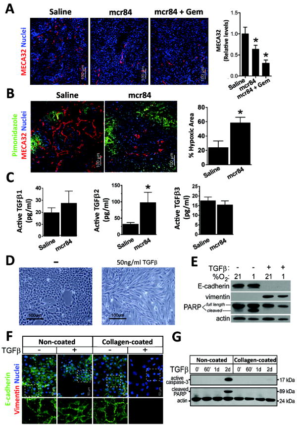
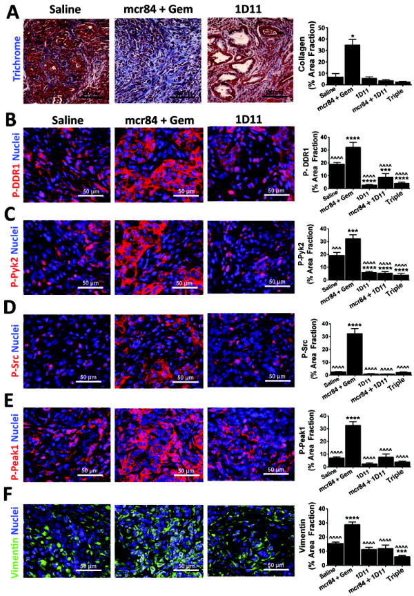
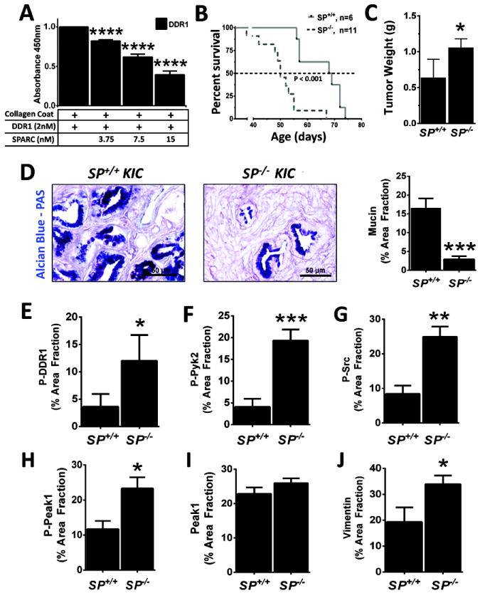
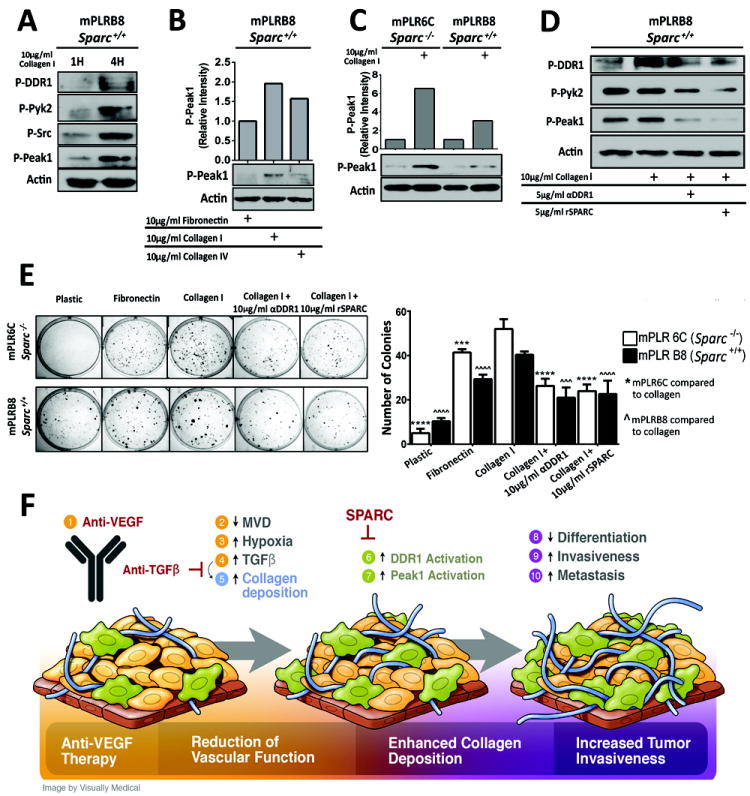
There is growing evidence that antiangiogenic therapy stimulates cancer cell invasion and metastasis. However, the underlying molecular mechanisms responsible for these changes have not been fully defined. Here, we report that anti-VEGF therapy promotes local invasion and metastasis by inducing collagen signaling in cancer cells. We show that chronic VEGF inhibition in a genetically engineered mouse model of pancreatic ductal adenocarcinoma (PDA) induces hypoxia, a less differentiated mesenchymal-like tumor cell phenotype, TGF-β expression, and collagen deposition and signaling. In addition, we show that collagen signaling is critical for protumorigenic activity of TGF-β in vitro. To further model the impact of collagen signaling in tumors, we evaluated PDA in mice lacking Sparc, a protein that reduces collagen binding to cell surface receptors. Importantly, we show that loss of Sparc increases collagen signaling and tumor progression. Together, these findings suggest that collagen actively promotes PDA spread and that enhanced disease progression associated with anti-VEGF therapy can arise from elevated extracellular matrix-mediated signaling.


Conflict of interest statement

Conflict of Interest Statement: R. A. Brekken is a consultant for, has equity interest in, and is a recipient of a sponsored research grant from Peregrine Pharmaceuticals and Affitech AS. R. A. Brekken is also an author of a patent on technology that was used to develop the antibody r84 by Peregrine Pharmaceuticals and Affitech. Peregrine and Affitech did not participate in the planning, execution, or interpretation of the experiments.

Figures

Figure 1
Figure 1. Activated endothelial cells are present in KIC PDA tumors
(A) KIC tumors exhibit abnormal vascular architecture. Wild-type (WT), and 4 and 7 week KIC samples were stained for a blood vessel marker (endomucin) and a pericyte marker (NG2). Quantification of microvessel density is displayed in (B). (C) Vessels undergo angiogenic stress as seen by increased vessel sprouting (Endomucin) in KIC tissues. (D) Endothelial cell activation in KIC PDA. WT pancreas and KIC PDA were stained for PCNA and endomucin. Inset in right panel highlights double-stained proliferating endothelial cells. Quantification of proliferating endothelial cells in KIC PDA is presented in (D). (E) Elevated expression of VEGF in PDA. VEGF expression in pancreata collected from 7 week old WT and KIC mice was assessed by qPCR. Error bars represent SEM (*, p < 0.05 vs WT; ***, p < 0.0005 vs WT). One way ANOVA with Tukey’s MCT.
Figure 2
Figure 2. VEGF-blockade restricts tumor growth and promotes survival of KIC mice
KIC mice were treated with saline (n=10), mcr84 (n=10), or mcr84 + Gem (n=11) from 4-8 weeks of age, after which pancreases were harvested (A-C). (A) mcr84 blocks the replacement of normal pancreatic parenchyma with PDA. Tissue sections from the indicated groups were stained for amylase, a marker of exocrine pancreas. Tumors in mcr84 and mcr84 plus Gem treated mice are circumscribed by a dotted line. Quantitation of amylase content is presented in (B). (C) mcr84 controls primary PDA growth. Mean pancreas weight was used a surrogate measurement of primary tumor burden. (D) Mice in a survival study were treated with saline, mcr84, or mcr84 + Gem from 4 weeks until moribund. (E) Metastasis to the liver was quantified by qPCR for recombined Cdkn2a allele (n=6-7 animals/group). (F) mcr84-treated KIC tumors exhibit a less differentiated phenotype as documented by H&E histology. (G) Collagen content is elevated after anti-VEGF therapy. Tumors from the survival study were stained with Masson’s trichrome and collagen was quantified and is presented in right panel. (H) VEGF-blockade enhances EMT. Tumors from the survival study were stained for E-cadherin, Zeb1, and vimentin. Insets demonstrate glandular E-cadherin staining, and nuclear Zeb1 staining in non-glandular tumor cells. Positive staining area was quantified and is presented in panels adjacent the micrographs. Error bars represent SEM. (*, p < 0.05 vs saline; **, p < 0.005 vs saline; ***, p < 0.0005 vs saline). One way ANOVA with Tukey’s MCT.
Figure 3
Figure 3. VEGF-blockade induces hypoxia and elevates TGFβ expression
(A) Microvessel density in tumors from KIC mice treated with saline, mcr84, mcr84 + Gem was determined by immunohistochemistry with MECA32 (red). Quantification of MVD is shown. (B) 6 week old KIC mice were treated with saline or mcr84 for one week. Animals were sacrificed 1 hour after pimonidazole injection (iv). Tissue was stained for endothelial cells (MECA32, red) and pimonidazole adducts (Hypoxyprobe, green). Hypoxic area was quantified and is presented in right panel. (C) Lysates from tumors harvested from saline and mcr84 treated KIC animals were assessed for active TGFβ proteins by ELISA. The level of TGFβ2 was elevated significantly by blockade of VEGF with mcr84. (D) Stimulation of primary KIC cells with TGFβ for 48 hours resulted in morphological changes. (E) TGFβ stimulation promoted loss of E-cadherin, gain of vimentin expression and protection from hypoxia-induced apoptosis. (F, G) Collagen protects PDA cells from TGFβ-induced apoptosis. Murine KIC-derived PDA cells (clone 3B) were plated on plastic or collagen-coated surfaces and treated with 10 ng/ml TGFβ1 for 72 hrs or the indicated time. (F) Cells were examined by immunocytochemistry for E-cadherin (green) and vimentin (red). Dashed insets are enlarged and presented beneath each panel. (G) Cell lysates were probed for active caspase-3 and cleavage of the caspase-3 substrate PARP. Actin was used as a loading control. 1d = 24 hours; 2d = 48 hours. Error bars represent SEM (*, p < 0.05 vs saline)
Figure 4
Figure 4. Collagen promotes PDA cell survival and EMT
KIC mice were treated with saline (n=4), mcr84 + Gem (n=5), 1D11 (n=6), 1D11+mcr84 (n=6), or the triple combination (n=8) in the context of a survival study. Pancreatic tissue was harvested and subjected to Masson’s Trichrome (A) or immunohistochemistry for phosphorylated Ddr1 (B, p-DDR1), phosphorylated Pyk2 (C, P-Pyk2), phosphorylated Src (D, P-Src), phosphorylated Peak1 (E, P-Peak1) or vimentin (F). Quantification of signal intensity for each target in each treatment group is shown. Error bars represent SEM (*, p < 0.05 vs saline; ***, p < 0.0005 vs saline; ˆˆˆ, p < 0.0005 vs mcr84+gem; ****, p < 0.00005 vs saline; ˆˆˆˆ, p < 0.00005 vs mcr84+gem). One way ANOVA with Tukey’s MCT.
Figure 5
Figure 5. Sparc attenuates PDA progression by reducing collagen signaling
(A) SPARC blocks interaction between DDR1 and collagen. The binding of 2 nM DDR1/Fc to collagen I was determined by ELISA with collagen I being the solid phase. The binding to DDR1/Fc was detected via the Fc tag in triplicate. Increasing concentrations of recombinant human SPARC reduced the binding DDR1/Fc to collagen I. Error bars represent SEM (****, p < 0.00005 vs DDR1). One way ANOVA with Tukey’s MCT. (B) Decreased survival of Sparc-/-:KIC mice. Sparc-/- were crossed with KIC mice to generate Sparc+/-:KIC animals which were crossed with each other to generate KIC (SP+/+) and Sparc-/-:KIC (SP-/-) animals. Survival of these animals is shown. (C) Mean pancreas/tumor weight in Sparc+/+:KIC and Sparc-/-:KIC animals. (D) Alcian blue-PAS histology of tumors from Sparc+/+:KIC and Sparc-/-:KIC animals. (E-J) Expression of mediators of collagen signaling in murine PDA. Tumor was harvested from moribund KIC and Sparc-/-:KIC mice, sectioned and stained for phosphorylated Ddr1 (E, P-DDR1), phosphorylated Pyk2 (F, P-Pyk2), phosphorylated Src (G, P-Src), phosphorylated Peak1 (H, P-Peak1), total Peak1 (I), and Vimentin (J). Error bars represent SEM (*, p < 0.05 vs SP+/+; **, p < 0.005 vs SP+/+; ***, p < 0.0005 vs SP+/+; ****, p < 0.00005 vs SP+/+).
Figure 6
Figure 6. PDA cells express collagen receptors and Sparc
(A) mPLRB8 cells were plated on fibronectin and stimulated with soluble collagen I (10 μg/ml) for 1 or 4 hours. Lysates were probed for the indicated targets by western blotting. (B) mPLRB8 cells were stimulated for 4 hours with fibronectin, collagen I or collagen IV and the level of active Peak1 was determined by western blotting. (C) Activation of Peak1 was determined in Sparc-/- (mPLR6C) and Sparc+/+ (mPLRB8) KIC cells after 4 hours of collagen I stimulation. (D) Blockade of DDR1 reduces Peak1 activation. Sparc+/+:KIC cell line (B8) plated on 5 μg/ml fibronectin. Cells were stimulated with no treatment, collagen I, collagen I + anti-DDR1 (5 μg/ml) or collagen I + rSPARC (5 μg/ml). Peak1 activation was measured after 24 hours of stimulation. (E) Functional consequences of Ddr1 blockade. Sparc-/-:KIC clone (mPLR6C) and Sparc+/+:KIC clone (mPLRB8) were plated on plastic, 10 μg/ml fibronectin, or 10 μg/ml collagen I in the presence or absence of Ddr1 inhibitors (DDr1 blocking antibody or recombinant SPARC) and colony formation was determined. (F) Schematic displaying the proposed consequences of anti-VEGF therapy (1), which include reduced vascular function (2-4), enhanced collagen deposition (5-7) and increased tumor invasion (8-10). Epithelial like tumor cells (gold color) adopt a mesenchymal-like phenotype (olive color), a process that is increased by therapy-induced hypoxia and elevated collagen signaling. Error bars represent SEM (***, p < 0.0005 vs mPLR6C; ˆˆˆ, p < 0.0005 vs mPLRB8; ****, p < 0.00005 vs mPLR6C; ˆˆˆˆ, p < 0.00005 vs mPLRB8). One way ANOVA with Tukey’s MCT.

References

    1. Folkman J. Tumor angiogenesis: therapeutic implications. N Engl J Med. 1971;285:1182–6. - PubMed
    1. Roskoski R., Jr Vascular endothelial growth factor (VEGF) signaling in tumor progression. Crit Rev Oncol Hematol. 2007;62:179–213. - PubMed
    1. Hanahan D, Weinberg RA. Hallmarks of cancer: the next generation. Cell. 2011;144:646–74. - PubMed
    1. Shojaei F. Anti-angiogenesis therapy in cancer: current challenges and future perspectives. Cancer Lett. 2012;320:130–7. - PubMed
    1. Welti J, Loges S, Dimmeler S, Carmeliet P. Recent molecular discoveries in angiogenesis and antiangiogenic therapies in cancer. J Clin Invest. 2013;123:3190–200. - PMC - PubMed

Publication types

MeSH terms