

A perspective on the role of the extracellular matrix in progressive retinal degenerative disorders
- PMID: 24346621
- PMCID: PMC4587794
- DOI: 10.1167/iovs.13-13536
A perspective on the role of the extracellular matrix in progressive retinal degenerative disorders
Abstract
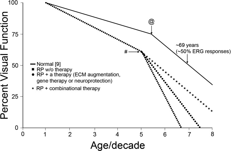
Progressive inherited retinal degenerative disorders (PIRDDs) are the leading cause of blindness in developed countries, with AMD and RP constituting the majority of PIRDDs. Currently, over 8 million Americans have PIRDDs, and that number is estimated to drastically increase by the end of this decade. Although a mutant protein is expressed starting early during retinal development in patients with PIRDDs, symptoms of retinal degeneration do not manifest until much later. Historically, research has focused on understanding the role a mutation has in the function of a protein and what role the mutant protein has in the disease process. However, it remains unknown why the disease, irrespective of the mutation, manifests clinically much later in life, while cellular indicators of disease (e.g., accumulation of toxic protein products and cell death) occur throughout early and middle life. Herein, we propose that there exists a time point at which the degenerative process is accelerated, leading to the appearance of clinical symptoms. This point is defined by structural disruptions of the extracellular matrix (ECM). Death of a critical number of ECM-maintaining mutant protein-expressing retinal cells contributes to that break point in the degenerative process. Therefore, it is important to understand the changes occurring at the ECM during PIRDDs and to take that into account when therapeutic approaches are designed.
Keywords: aging; extracellular matrix; retinal degeneration.
Figures
References
-
- Shastry BS. Retinitis pigmentosa and related disorders: phenotype of rhodopsin and peripherin/rds mutations. Am J Med Genet. 1994; 52: 467–474. - PubMed
-
- Roth S. Endogenous neuroprotection in the retina. Brain Res Bull. 2004; 62: 461–466. - PubMed
-
- Mecham RP. Overview of extracellular matrix. Curr Protoc Cell Biol. 2012; chap 10:unit 10.1. - PubMed
Publication types
MeSH terms
Grants and funding
LinkOut - more resources
Full Text Sources
Other Literature Sources
Medical
