. 2014 Mar;34(3):433-40.
doi: 10.1038/jcbfm.2013.213. Epub 2013 Dec 18.

The Wnt/planar cell polarity signaling pathway contributes to the integrity of tight junctions in brain endothelial cells

Affiliations

The Wnt/planar cell polarity signaling pathway contributes to the integrity of tight junctions in brain endothelial cells

Cédric Artus et al. J Cereb Blood Flow Metab. 2014 Mar.

Abstract

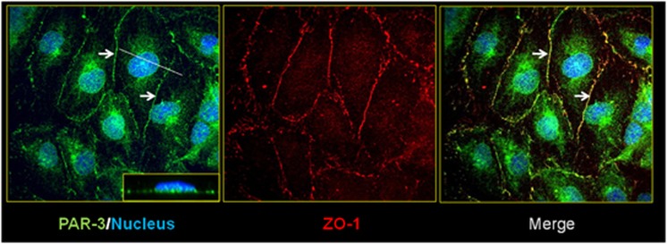
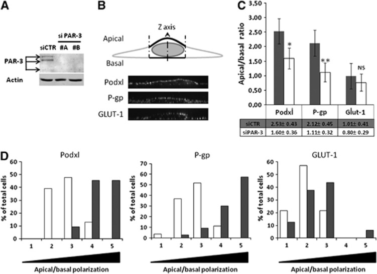
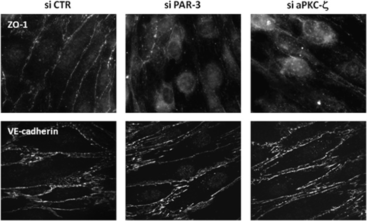
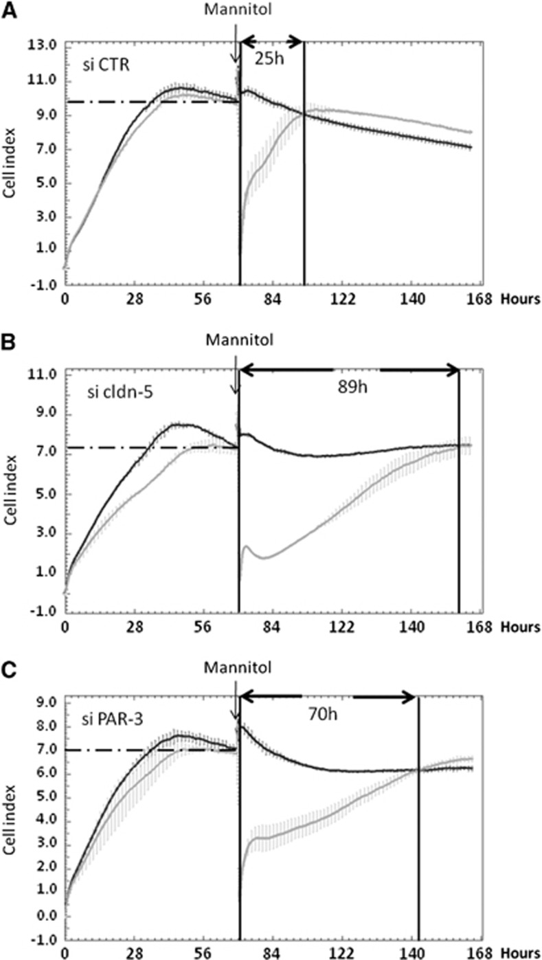
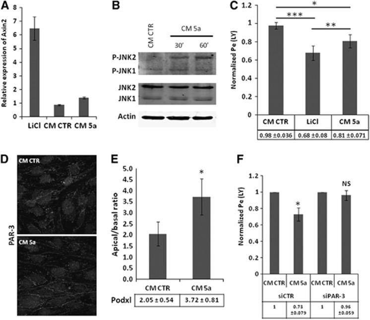
Wnt morphogens released by neural precursor cells were recently reported to control blood-brain barrier (BBB) formation during development. Indeed, in mouse brain endothelial cells, activation of the Wnt/β-catenin signaling pathway, also known as the canonical Wnt pathway, was shown to stabilize endothelial tight junctions (TJs) through transcriptional regulation of the expression of TJ proteins. Because Wnt proteins activate several distinct β-catenin-dependent and independent signaling pathways, this study was designed to assess whether the noncanonical Wnt/Par/aPKC planar cell polarity (PCP) pathway might also control TJ integrity in brain endothelial cells. First we established, in the hCMEC/D3 human brain endothelial cell line, that the Par/aPKC PCP complex colocalizes with TJs and controls apicobasal polarization. Second, using an siRNA approach, we showed that the Par/aPKC PCP complex regulates TJ stability and reassembling after osmotic shock. Finally, we provided evidence that Wnt5a signals in hCMEC/D3 cells through activation of the Par/aPKC PCP complex, independently of the Wnt canonical β-catenin-dependent pathway and significantly contributes to TJ integrity and endothelial apicobasal polarity. In conclusion, this study suggests that the Wnt/Par/aPKC PCP pathway, in addition to the Wnt/β-catenin canonical pathway, is a key regulator of the BBB.


Figures

Figure 1
Figure 1
Par/aPKC planar cell polarity complex is expressed in hCMEC/D3 cells and colocalized with ZO-1. HCMEC/D3 cells grown at confluence for 6 days were fixed and permeabilized. Double immunofluorescence labeling was performed with a ZO-1 polyclonal antibody and PAR-3 monoclonal antibody. Nuclei were labeled with 4′,6-diamidino-2-phenylindole. Inset in right panel xz plan, which represent PAR-3 staining.
Figure 2
Figure 2
The Par/aPKC planar cell polarity complex controls hCMEC/D3 cells polarization. (A) Immunoblot analysis of PAR-3 and actin expression. hCMEC/D3 cells were treated with control small interfering RNA (si CTR) or two distinct siRNAs against PAR-3 (#A, #B). Cells were grown at confluence and whole-cell lysates were generated. Proteins were separated on sodium dodecyl sulfate polyacrylamide gel electrophoresis gel followed by immunoblotting with anti-PAR-3 or anti-actin monoclonal antibodies. (B) Cell membranes were stained using CM-dil then cells were fixed and permeabilized. Immunostaining was performed using Podxl or P-gp monoclonal antibodies or a GLUT-1 polyclonal antibody. Nuclei were labeled with DAPI. Membranous immunofluorescence intensity was evaluated along cells Z-axis above and below the nucleus using ImageJ software and curve was generated using KaleidaGraph software. Apical or basal fluorescence intensity was calculated by integrating area under apical or basal peak defined by membrane labeling with CM-dil. (C) HCMEC/D3 cells treated with control siRNA (gray bars) or PAR-3 siRNA (#A) (white bars) were grown at confluence for 6 days on culture inserts. Mean apical/basal fluorescence intensity of 40 different cells from three independent experiments was calculated. Indicated P values were obtained using Student's t-test: *P<0.005; **P<0.001; NS, not significant. (D) Cell repartition in percent between apical/basal ratio range divided in five equal categories (gray: siCTR; white: siPAR-3). Category 1 includes cells with lower apical/basal ratio (0 to 0.5) to category 5, which includes cells with higher apical/basal ratio (2 to 2.5).
Figure 3
Figure 3
Par/aPKC planar cell polarity complex knockdown increases hCMEC/D3 permeabilty to Lucifer Yellow (LY). (A) Immunoblot analysis of atypical protein kinase C-ζ (aPKC-ζ) and actin expression. HCMEC/D3 cells were treated with control small interfering RNA (si CTR) or two distinct siRNAs against aPKC-ζ. Cells were grown at confluence and whole-cell lysates were generated. Proteins were separated on sodium dodecyl sulfate polyacrylamide gel electrophoresis gel followed by immunoblotting with anti-aPKC-ζ or anti-actin monoclonal antibodies. aPKC-ζ immunoblot quantification using ImageJ software, relative expression of aPKC-ζ normalized to actin level (lower panel). (B) HCMEC/D3 cells were treated with control siRNA or PAR-3 siRNA (#A) or two distinct aPKC-ζ siRNA (#1, #2) and grown at confluence for 6 days on culture inserts. Permeability assays to LY were performed and the permeability coefficient (Pe) was quantified. Results are indicated as permeability ratio, normalized relative to the Pe of cells treated with control siRNA. The basal permeability level was 1.42±0.16.10−3 cm/minute. Results are means±s.d. for three independent experiments. Indicated P values were obtained using Student's t-test. *P<0.02; **P<0.005.
Figure 4
Figure 4
Par/aPKC planar cell polarity complex knockdown destabilizes tight junction complexes. HCMEC/D3 cells were treated with control small interfering RNAs (si CTR), PAR-3 siRNA (A) or atypical protein kinase C-ζ (aPKC-ζ) siRNA (#2), and grown at confluence for 6 days on culture inserts. Cells were fixed and permeabilized. Immunofluorescence labeling was performed with a ZO-1 monoclonal antibody and VE-cadherin (VE-cad) polyclonal antibody.
Figure 5
Figure 5
PAR-3 knockdown delays tight junction complex reassembling after 1 mol/L mannitol treatment. HCMEC/D3 cells treated with control (A), claudin-5 (cldn-5) (B) or PAR-3 (C) small interfering RNAs were grown on a 96-well E-plate. Impedance measurement was monitored in real time by xCELLigence system and curves were generated using the RTCA Software. After 72 hours, cells were treated with 1 mol/L mannitol. After another 30 minutes, medium was changed to normal growth medium for a recovery period of up to 4 days. Black curve: untreated cells; light gray curve: 1 mol/L mannitol-treated cells. Results are mean cell index (CI) values±s.d. (n=3 wells) from one representative experiment out of three independent experiments.
Figure 6
Figure 6
Wnt5a-triggered control of hCMEC/D3 permeability is mediated by Par/aPKC planar cell polarity complex. (A) HCMEC/D3 cells were incubated with EBM-2 medium or with EBM-2 containing 10 mmol/L lithium chloride (LiCl), 40% of control (CM CTR) or Wnt5a (CM 5a)-conditioned medium and grown at confluence for 6 days on culture inserts. Total RNA extracts were obtained using RNA kit extraction. Reverse transcription was performed and Axin2 messenger RNA expression was evaluated by quantitative real time polymerase chain reaction using LightCycler 480 device. Results are visualized as relative expression compared with EBM-2 medium. (B) Immunoblot analysis of JNK phosphorylation and actin expression. HCMEC/D3 cells grown at confluence for 6 days were treated for 30 minutes or 1 hour with EBM-2 completed with 40% of CM CTR or CM 5a. Whole-cell lysates were generated. Proteins were separated on sodium dodecyl sulfate polyacrylamide gel electrophoresis gel followed by immunoblotting with anti-Phospho-JNK polyclonal antibody, anti-JNK monoclonal antibody or anti-actin monoclonal antibody. (C) HCMEC/D3 cells were incubated with EBM-2 medium or with EBM-2 containing 10 mmol/L LiCl, 40% of CM CTR or CM 5a-conditioned medium and grown at confluence for 6 days on culture inserts. Permeability assays to Lucifer Yellow (LY) were performed and the permeability coefficient was quantified. Results are expressed as permeability ratio, normalized relative to the permeability coefficient for EBM-2 cultured cells. The basal permeability level was 1.8±0.1.10−3 cm/minute. Results are means±s.d. for seven independent experiments. Indicated P values were obtained using Student's t-test. *P<0.01; **P<0.003, ***P<0.002. (D) HCMEC/D3 cells were treated with EBM-2 complement with 40% of CM CTR or CM 5a and grown at confluence for 6 days on culture inserts. Then cells were fixed and permeabilized. Immunofluorescence labeling was performed with a PAR-3 polyclonal antibody. (E) HCMEC/D3 cells were treated with EBM-2 completed with 40% of CM CTR or CM 5a and grown at confluence for 6 days on culture inserts. Mean apical/basal fluorescence intensity of 40 different cells from at least three independent experiments is shown. Indicated P value was obtained using Student's t-test: *P<0.0001. (F) HCMEC/D3 cells treated with control siRNA or PAR-3 siRNA (#A) were grown at confluence for 6 days in EBM-2 medium completed with 40% of CM CTR or CM 5a on culture inserts. Permeability assays to LY were performed and the permeability coefficient was quantified. Results are expressed as permeability ratio, normalized relative to the permeability coefficient for CM CTR. The basal permeability level was 1.65±0.2.10−3 cm/minute. Results are means±s.d. for five independent experiments. Indicated P values were obtained using Student's t-test. *P<0.02; NS, not significant.

References

    1. Abbott NJ, Patabendige AA, Dolman DE, Yusof SR, Begley DJ. Structure and function of the blood-brain barrier. Neurobiol Dis. 2010;37:13–25. - PubMed
    1. Tsukita S, Furuse M, Itoh M. Multifunctional strands in tight junctions. Nat Rev Mol Cell Biol. 2001;2:285–293. - PubMed
    1. Dragsten PR, Blumenthal R, Handler JS. Membrane asymmetry in epithelia: is the tight junction a barrier to diffusion in the plasma membrane. Nature. 1981;294:718–722. - PubMed
    1. Furuse M, Hirase T, Itoh M, Nagafuchi A, Yonemura S, Tsukita S. Occludin: a novel integral membrane protein localizing at tight junctions. J Cell Biol. 1993;123:1777–1788. - PMC - PubMed
    1. Martin-Padura I, Lostaglio S, Schneemann M, Williams L, Romano M, Fruscella P, et al. Junctional adhesion molecule, a novel member of the immunoglobulin superfamily that distributes at intercellular junctions and modulates monocyte transmigration. J Cell Biol. 1998;142:117–127. - PMC - PubMed

Publication types

MeSH terms

LinkOut - more resources