. 2014 Mar;7(2):172-81.
doi: 10.1007/s12265-013-9526-4. Epub 2013 Dec 18.

Identification of streptococcal m-protein cardiopathogenic epitopes in experimental autoimmune valvulitis

Affiliations

Identification of streptococcal m-protein cardiopathogenic epitopes in experimental autoimmune valvulitis

Christine A Kirvan et al. J Cardiovasc Transl Res. 2014 Mar.

Abstract

The M protein of rheumatogenic group A streptococci induces carditis and valvulitis in Lewis rats and may play a role in pathogenesis of rheumatic heart disease. To identify the epitopes of M5 protein that produce valvulitis, synthetic peptides spanning A, B, and C repeat regions contained within the extracellular domain of the streptococcal M5 protein were investigated. A repeat region peptides NT4, NT5/6, and NT7 induced valvulitis similar to the intact pepsin fragment of M5 protein. T cell lines from rats with valvulitis recognized M5 peptides NT5/6 and NT6. Passive transfer of an NT5/6-specific T cell line into naïve rats produced valvulitis characterized by infiltration of CD4+ cells and upregulation of VCAM-1, while an NT6-specific T cell line did not target the valve. Our new data suggests that M protein-specific T cells may be important mediators of valvulitis in the Lewis rat model of rheumatic carditis.


Figures

Figure 1 (A-E)
Figure 1 (A-E)
Histopathologic features of valvulitis in Lewis rat heart sections stained with hematoxylin and eosin. A. Valvulitis is shown in rats immunized with group A streptococcal pepM5 (magnification, ×20) with the presence of mononuclear cell infiltrates directly under the valvular endothelium and in deeper valve tissue (small arrow) and inset (magnification,×200). Areas of edema are seen throughout the valve tissue (large arrows). Unaffected myocardium is shown (dashed arrow). B. Animals that received adjuvant alone had no detectable histopathologic lesions or edema (magnification,×20). Valves from rats immunized with pepM5 demonstrated the presence of both CD4+ (C) and CD8+ (D) T cells in comparison to the adjuvant control (E) (magnification,×40).
Figure 2 (A-F)
Figure 2 (A-F)
Induction of valvulitis and cellular infiltration in hematoxylin- and eosin-stained heart valves from Lewis rats immunized with group A streptococcal M5 peptide groups(Table 2). A. Lewis rats immunized with M5 peptide group 1 (NT1-4/5) revealed the presence of mononuclear cells (arrows) within mitral valve (magnification,×40) in relation to surface endothelial cells (dashed arrow). B. Cellular infiltrates (arrows) in valvular tissue from group A streptococcal M5 peptide group 1-immunized animals (magnification,×400). No cellular infiltrates were observed in the myocardium. C and D. Group 2 M5 peptides (NT 5-8) induced mononuclear cell invasion (arrows, magnification,×40 and×400) and provoked a strong inflammatory response observed by the presence of edema in the valve (E, large arrows). F. In contrast, group 3 peptides (B1A-B2B3A) induced small focal lesions within the myocardium with mononuclear cell infiltrates.
Figure 3 (A-D)
Figure 3 (A-D)
Identification of specific streptococcal M5 peptides that induced valvulitis and myocarditis from the A repeat region of pep M5. Peptide NT4 induced both valvulitis (A) and myocarditis (B) similar to group A streptococcal pepM5 protein. Valvulitis produced by peptide NT5/6 (C) is characterized by in invasion of both small mononuclear cells and macrophages. Prominent mononuclear cell infiltrates are present in streptococcal M5 peptide NT7-induced valvulitis (D). Small arrows indicate invading mononuclear cells and large arrows denote the presence of edema within the valvular tissues. Valves from NT7 immunized rats revealed the presence of Anitschkow-like cells (dashed arrow). Magnification at×40 for all tissues.
Figure 4 (A-D)
Figure 4 (A-D)
Passive transfer of streptococcal M protein specific T cell lines produced valvulitis in naïve Lewis rats. NT5/6-specific T cell line p1.14 migrated to the mitral valve of naïve Lewis rats (A and B) in contrast to NT6-specific T cell line p1.8, which did not infiltrate heart valves (C). T cell line 1.17, which was M5-specific and recognized M5 peptides B2 and B3A, migrated to cardiac valves (D), similar to p1.14 (small arrows indicate endothelium). Large arrows indicate mononuclear cells (A-D). Magnification at×40 for all tissues.
Figure 5 (A-D)
Figure 5 (A-D)
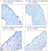
A. Cardiac tissues illustrate infiltration of CD4+ cells and detectable upregulation of VCAM-1 (C) after passive transfer of NT5/6-specific T cell line p1.14. Passive transfer of NT6-specific T cell line 1.8 did not reveal the presence of CD4+ cells (B) or upregulation of VCAM-1 (D) in the valve. Magnification,×40.
Figure 6
Figure 6
Amino acid sequence of alignment of streptococcal M5 protein amino acid sequence recognized by Lewis rat M5 specific T cell lines associated with passive transfer of valvulitis compared with the streptococcal M5 protein amino acid sequence recognized by an intralesional T cell clone isolated from valve in human rheumatic carditis [24].

References

    1. Stollerman GH. Rheumatic fever in the 21st century. Clin Infect Dis. 2001;33(6):806–14. - PubMed
    1. Stollerman GH. Rheumatic Fever. Lancet. 1997;349:935–942. - PubMed
    1. Cunningham MW. Pathogenesis of Acute Rheumatic Fever. In: Stevens DS, Kaplan EH, editors. Streptococcal Diseases. first. New York, NY: Oxford Press; 2000. pp. 102–132.
    1. Cunningham MW. Streptococcus and Rheumatic Fever. Current Opinion in Rheumatology. 2012;24:408–416. - PMC - PubMed
    1. Dajani AS. Guidelines for the diagnosis of rheumatic fever (Jones criteria, 1992 update) Journal of the American Medical Association. 1992;268:2069–2073. - PubMed

MeSH terms