

Granuloma faciale: a rare disease from a dermoscopy perspective
- PMID: 24346891
- PMCID: PMC3875968
- DOI: 10.1590/abd1806-4841.20132384
Granuloma faciale: a rare disease from a dermoscopy perspective
Abstract
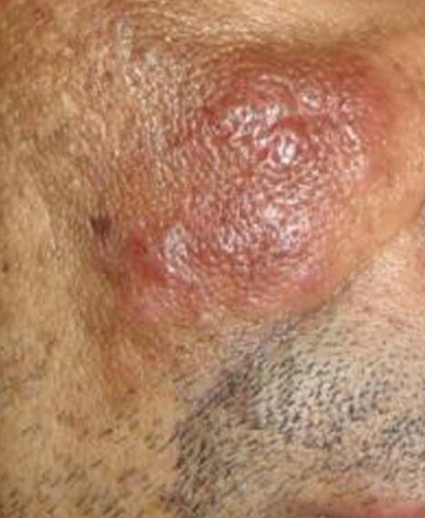
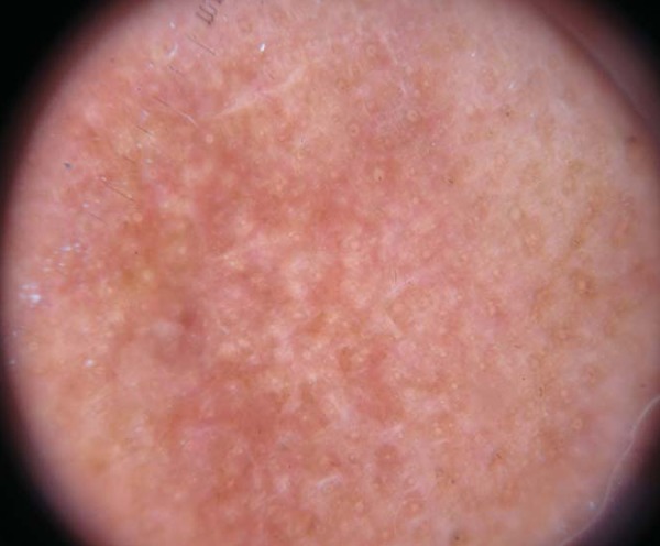
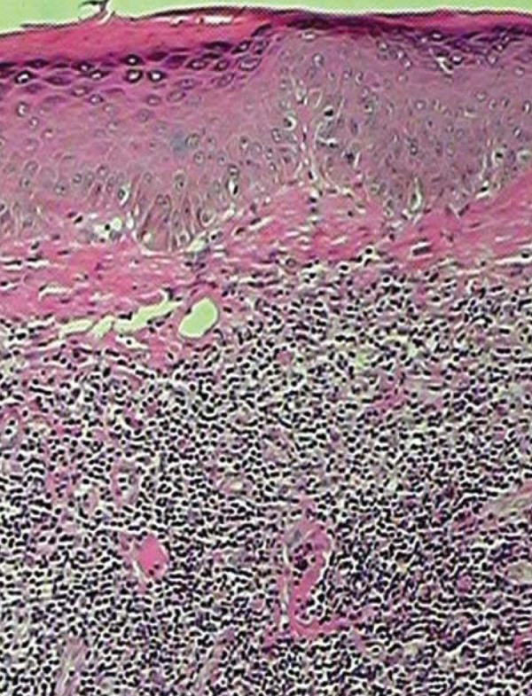
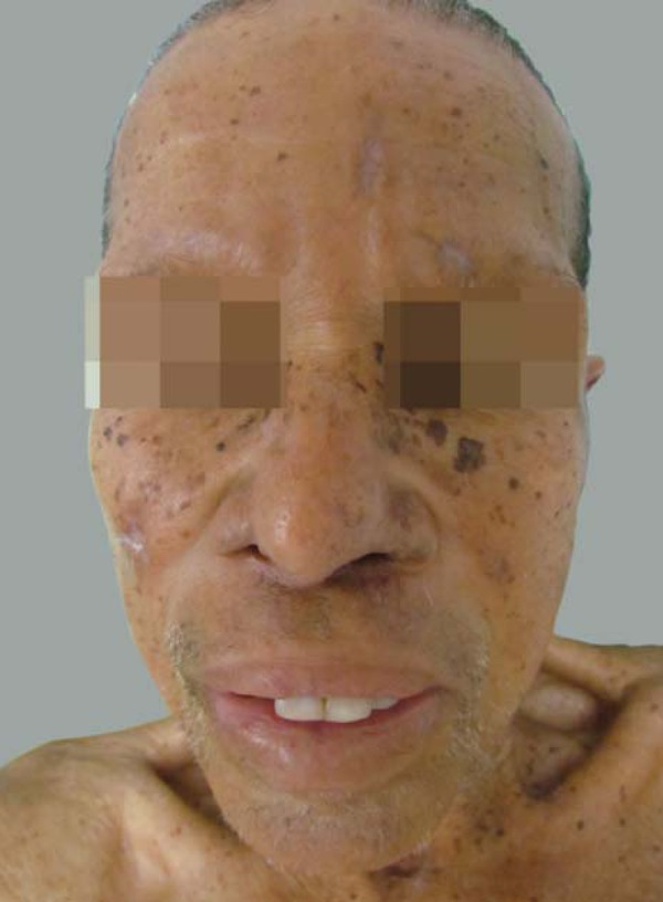
The granuloma faciale is a rare and benign skin disease of unknown etiology, characterized by chronic leukocitoclastic vasculitis. It is characterized by skin lesions predominantly facial whose course is chronic and slowly progressive. The diagnosis is based on clinical features, histopathology and, more recently, in dermoscopy. We describe the case of a male patient, 40 years old, with a sarcoid lesion on the malar site, whose histopathological examination revealed a mixed inflammatory infiltrate with presence of Grenz zone. Dermoscopy revealed a pink background with white striations. The definitive diagnosis is made by histopathologic evaluation, and dermatoscopy can be helpful. It is known to be resistant to therapy, oral medications, intralesional and surgical procedures are options.
O granuloma facial é doença cutânea rara e benigna, de etiologia desconhecida, caracterizado por vasculite leucocitoclástica crônica. Caracteriza-se por lesões cutâneas predominantemente faciais, tem curso crônico e lentamente progressivo. O diagnóstico é baseado na clínica, histopatologia e, mais recentemente, na dermatoscopia. Relatamos o quadro de um paciente masculino, 40 anos de idade, com lesão sarcoídea na face malar, cujo exame histopatológico revelou infiltrado inflamatório misto com presença de zona de Grenz. A dermatoscopia revelou um fundo rosado com estrias brancas. O diagnóstico definitivo é feito pela avaliação histopatológica, sendo que a dermatoscopia pode ser útil. É conhecida por ser resistente à terapêutica, sendo propostas medicações orais, intralesionais e procedimentos cirúrgicos.
Conflict of interest statement
Conflict of Interests: None.
Figures
References
-
- Caldarola G, Zalaudek I, Argenziano G, Bisceglia M, Pellicano R. Granuloma faciale: a case report on long-term treatment with topical tacrolimus and dermatoscopic aspects. Dermatol Ther. 2011;24:508–511. - PubMed
-
- Ito LM, Barros JF, Andrade R, Neves SRC. Granuloma faciale: case study. An Bras Dermatol. 1999;74:245–247.
-
- Marcoval J, Moreno A, Peyr J. Granuloma faciale: a clinicopathological study of 11 cases. J Am Acad Dermatol. 2004;51:269–273. - PubMed
-
- Ludwig E, Allam JP, Bieber T, Novak N. New treatment modalities for granuloma faciale. Br J Dermatol. 2003;149:634–637. - PubMed
-
- Thiyanaratnam J, Doherty SD, Krishnan B, Hsu S. Granuloma faciale: case report and review. Dermatol Online J. 2009;15:3. - PubMed
Publication types
MeSH terms
Substances
LinkOut - more resources
Full Text Sources
Other Literature Sources