

Prohibitins role in cellular survival through Ras-Raf-MEK-ERK pathway
- PMID: 24347342
- PMCID: PMC4413917
- DOI: 10.1002/jcp.24531
Prohibitins role in cellular survival through Ras-Raf-MEK-ERK pathway
Abstract
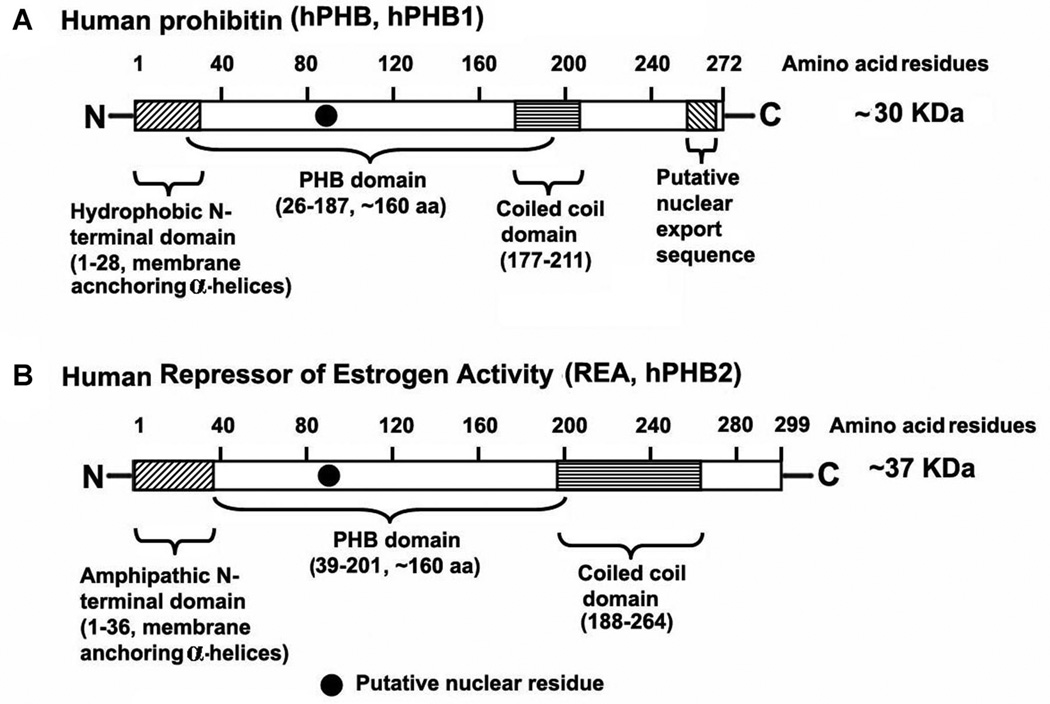
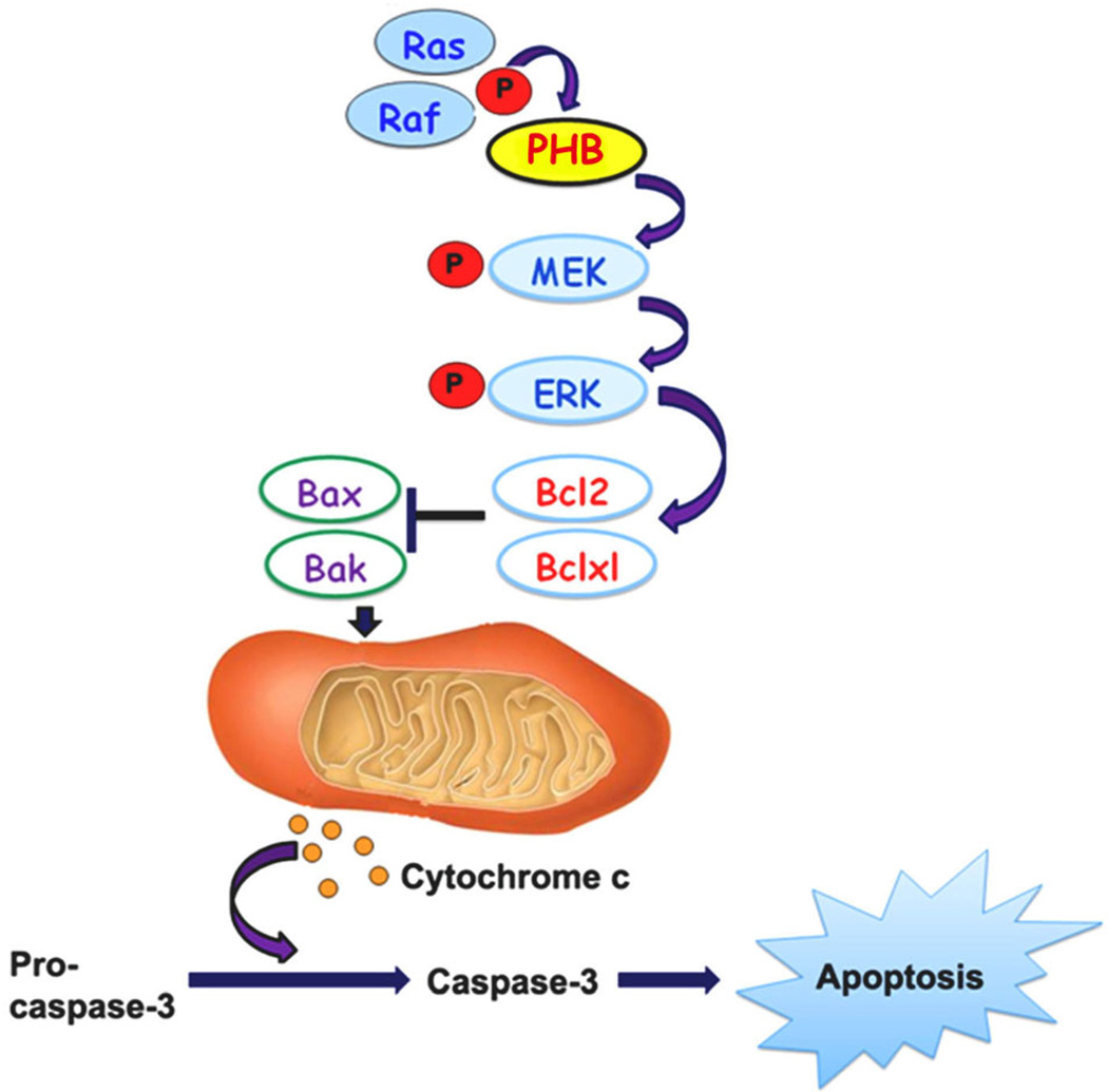
Prohibitins are members of a highly conserved protein family containing the stomatin/prohibitin/flotillin/HflK/C (SPFH) domain (also known as the prohibitin [PHB] domain) found in unicellular eukaryotes, fungi, plants, animals, and humans. Two highly homologous members of prohibitins expressed in eukaryotes are prohibitin (PHB; B-cell receptor associated protein-32, BAP-32) and prohibitin 2/repressor of estrogen receptor activity (PHB2, REA, BAP-37). Both PHB and REA/PHB2 are ubiquitously expressed and are present in multiple cellular compartments including the mitochondria, nucleus, and the plasma membrane. Multiple functions have been attributed to the mitochondrial and nuclear PHB and PHB2/REA including cellular differentiation, anti-proliferation, and morphogenesis. One of the major functions of the prohibitins are in maintaining the functional integrity of the mitochondria and protecting cells from various stresses. In the present review, we focus on the recent research developments indicating that PHB and PHB2/REA are involved in maintaining cellular survival through the Ras-Raf-MEK-Erk pathway. Understanding the molecular mechanisms by which the intracellular signaling pathways utilize prohibitins in governing cellular survival is likely to result in development of therapeutic strategies to overcome various human pathological disorders such as diabetes, obesity, neurological diseases, inflammatory bowel disease, and cancer.
© 2013 Wiley Periodicals, Inc.
Figures
References
-
- Ahn CS, Lee JH, Reum Hwang A, Kim WT, Pai HS. Prohibitin is involved in mitochondrial biogenesis in plants. Plant J. 2006;46:658–667. - PubMed
-
- Akepati VR, Müller EC, Otto A, Strauss HM, Portwich M, Alexander C. Characterization of OPA1 isoforms isolated from mouse tissues. J Neurochem. 2008;106:372–383. - PubMed
-
- Alexander C, Votruba M, Pesch UE, Thiselton DL, Mayer S, Moore A, Rodriguez M, Kellner U, Leo-Kottler B, Auburger G, Bhattacharya SS, Wissinger B. OPA1, encoding a dynamin-related GTPase, is mutated in autosomal dominant optic atrophy linked to chromosome 3q28. Nat Genet. 2000;26:211–215. - PubMed
-
- Altus MS, Wood CM, Stewart DA, Roskams AJ, Friedman V, Henderson T, Owens GA, Danner DB, Jupe ER, Dell’Orco RT, Keith McClung J. Regions of evolutionary conservation between the rat and human prohibitin-encoding genes. Gene. 1995;158:291–294. - PubMed
-
- Ande SR, Mishra S. Prohibitin interacts with phosphatidylinositol 3,4,5-triphosphate (PIP3) and modulates insulin signaling. Biochem Biophys Res Commun. 2009;390:1023–1028. - PubMed
Publication types
MeSH terms
Substances
Grants and funding
LinkOut - more resources
Full Text Sources
Other Literature Sources
Molecular Biology Databases
Research Materials
Miscellaneous
