. 2014 Sep 15;135(6):1381-9.
doi: 10.1002/ijc.28670. Epub 2014 May 8.

Comprehensive metagenomic analysis of glioblastoma reveals absence of known virus despite antiviral-like type I interferon gene response

Affiliations

Comprehensive metagenomic analysis of glioblastoma reveals absence of known virus despite antiviral-like type I interferon gene response

Érika Cosset et al. Int J Cancer. .

Abstract

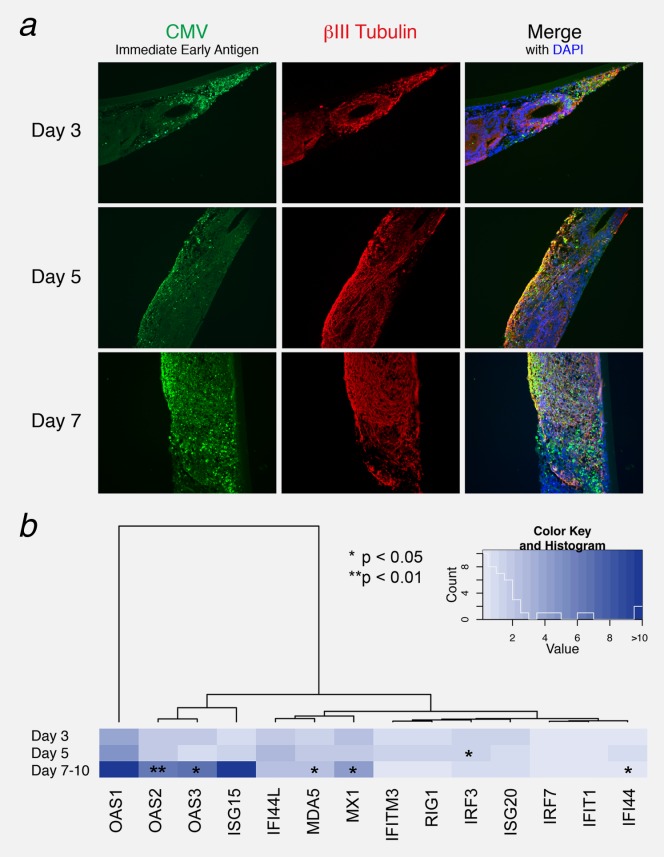
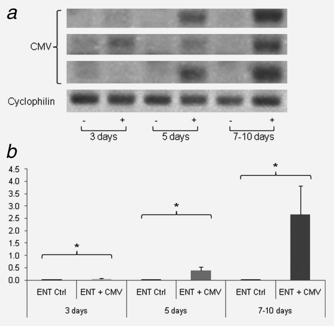
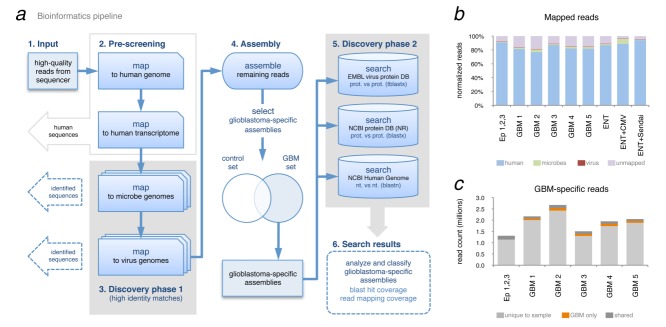
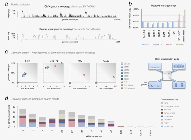
Glioblastoma is a deadly malignant brain tumor and one of the most incurable forms of cancer in need of new therapeutic targets. As some cancers are known to be caused by a virus, the discovery of viruses could open the possibility to treat, and perhaps prevent, such a disease. Although an association with viruses such as cytomegalovirus or Simian virus 40 has been strongly suggested, involvement of these and other viruses in the initiation and/or propagation of glioblastoma remains vague, controversial and warrants elucidation. To exhaustively address the association of virus and glioblastoma, we developed and validated a robust metagenomic approach to analyze patient biopsies via high-throughput sequencing, a sensitive tool for virus screening. In addition to traditional clinical diagnostics, glioblastoma biopsies were deep-sequenced and analyzed with a multistage computational pipeline to identify known or potentially discover unknown viruses. In contrast to the studies reporting the presence of viral signatures in glioblastoma, no common or recurring active viruses were detected, despite finding an antiviral-like type I interferon response in some specimens. Our findings highlight a discrete and non-specific viral signature and uncharacterized short RNA sequences in glioblastoma. This study provides new insights into glioblastoma pathogenesis and defines a general methodology that can be used for high-resolution virus screening and discovery in human cancers.

Keywords: antiviral type I interferon response; glioblastoma multiforme; high-throughput sequencing; metagenomic analysis; virus discovery.


Figures

Figure 1
Figure 1
IFN-related gene expression in GBM patient biopsies and other cancers. Expression levels of ISG20, ISG15, Mx1, OAS1, 2 and 3, IFITM3, IFIT1, IFI44, IFI44L, MDA-5, IRF7, STAT1 and RIG1 were detected by quantitative RT-PCR. The heat map depicts values of the fold increase expression of each gene normalized to two housekeeping genes (TBP and GAPDH). Hierarchical clustering illustrates the heterogeneity of gene expression profiles for each type of cancer. GBM biopsies that were deep-sequenced are marked with a red dot.
Figure 2
Figure 2
Infection of human brain-like tissue with CMV induces IFN-related gene expression. ENTs were infected with CMV and IFN-related gene expression was analyzed 3, 5 and 7–10 days post-infection. (a) CMV immediate early antigen (IEA) immunofluorescence detection. While little fluorescence was observed at 3 days, the entirety of the neural tissue (marked by beta3-Tubulin) was infected by 7–10 days post-infection. (b) Expression levels of type I IFN-related genes were analyzed by quantitative RT-PCR. The fold increase in expression of each gene was normalized to three housekeeping genes (TBP, ALAS1 and EEF1). Heatmap colors depict the fold increase in expression of each ENT + CMV relative to uninfected ENT at different times post-infection. Data are represented as mean (n = 3) ± SEM (*p < 0.05; **p < 0.01).
Figure 3
Figure 3
Nested PCR and qPCR of ENTs infected with CMV. ENTs were infected with CMV 3, 5 and 7 days post-infection. CMV was detected only in ENTs infected with the virus by Nested PCR (a) and semi-qPCR (b). Graphs depict the fold increase of CMV expression. Data are represented as mean (n = 4) ± SEM (*p < 0.05, “−” = non-infected ENT, “+” = infected ENT).
Figure 4
Figure 4
Metagenomic analysis of GBM deep-sequence data. (a) Bioinformatics pipeline developed to filter sequencing reads and discover known viruses (high identity matches step 3), assemble remaining reads and search for more distant similarities to virus (low-identity search step 5). (b) Proportion of mapped reads per sample, per organism. (c) Comparison of read-identity between GBM samples. Per sample, each sequence read with 100% identity to a read found in another sample is labeled as shared (among all samples including control), shared only in GBM samples or not shared (unique to that sample).
Figure 5
Figure 5
Virus detection in GBM deep-sequence data. (a) Pipeline validation and positive controls. Discovery phase 1 can capture genome-wide signatures of both DNA (CMV, maximum 110 X-fold coverage) and RNA (Sendai maximum 2,308 X-fold coverage) viruses. (b) Detected virus genomes shown as percentage of total non-human reads mapped to each genome. (c) Virus genome percent coverage and average depth of coverage from discovery phase 1 for the four detected viruses (PIV-5, phage phiX 174, CMV and Sendai virus). Refer to guide on right side for interpreting chart regions (I) low % genome coverage with little depth (number of reads mapped to genome), (II) low % coverage—great depth, (III) high % coverage—little depth and (IV) high % coverage—great depth. (d) Identity of assembled sequences found in two or more GBM specimens. After finding common sequences in GBM-specific assemblies (100% nucleotide sequence identity found in 2, 3, 4 or 5 GBM specimens), each assembly was Blasted against virus, nr and human sequence databases. Assemblies were classified according to databases in which they had significant matches; often an assembly had significant (e-value ≤0.001) matches in more than one database. Sample sets with no common sequences are not listed (e.g., [1,2,4], [2,3,4], etc.).
Figure 6
Figure 6
PCR validation of assembled contigs. The assembly process was verified by PCR followed by sequencing. The PCRs were performed on several biopsies: epileptic (Ep), Glioblastoma multiforme (GBM), breast carcinoma brain metastasis (BCBM) and astrocytoma grade II (AII). Amplified DNA products from all samples were visualized on agarose gels with ethidium bromide. PCR amplicons were then extracted and sequenced (DNA Genetic Analyzer 3130XL Applied Biosystems). (a) TIMP1 and (b) α-satellite DNA (α-satDNA) sequence identities were confirmed by NCBI BLAST.

References

    1. Louis DN, Ohgaki H, Wiestler OD, et al. The 2007 WHO classification of tumours of the central nervous system. Acta Neuropathol. 2007;114:97–109. - PMC - PubMed
    1. Stupp R, Mason WP, van den Bent MJ, et al. Radiotherapy plus concomitant and adjuvant temozolomide for glioblastoma. N Engl J Med. 2005;352:987–996. - PubMed
    1. Schwartzbaum JA, Fisher JL, Aldape KD, et al. Epidemiology and molecular pathology of glioma. Nat Clin Pract Neurol. 2006;2:494–503; quiz 1 p following 16. - PubMed
    1. Ohgaki H, Kleihues P. Epidemiology and etiology of gliomas. Acta Neuropathol. 2005;109:93–108. - PubMed
    1. Kouhata T, Fukuyama K, Hagihara N, et al. Detection of simian virus 40 DNA sequence in human primary glioblastomas multiforme. J Neurosurg. 2001;95:96–101. - PubMed

Publication types