. 2014 Jan 14;111(2):763-8.
doi: 10.1073/pnas.1311520111. Epub 2013 Dec 17.

Spontaneous slow replication fork progression elicits mitosis alterations in homologous recombination-deficient mammalian cells

Affiliations

Spontaneous slow replication fork progression elicits mitosis alterations in homologous recombination-deficient mammalian cells

Therese Wilhelm et al. Proc Natl Acad Sci U S A. .

Abstract

Homologous recombination deficient (HR(-)) mammalian cells spontaneously display reduced replication fork (RF) movement and mitotic extra centrosomes. We show here that these cells present a complex mitotic phenotype, including prolonged metaphase arrest, anaphase bridges, and multipolar segregations. We then asked whether the replication and the mitotic phenotypes are interdependent. First, we determined low doses of hydroxyurea that did not affect the cell cycle distribution or activate CHK1 phosphorylation but did slow the replication fork movement of wild-type cells to the same level than in HR(-) cells. Remarkably, these low hydroxyurea doses generated the same mitotic defects (and to the same extent) in wild-type cells as observed in unchallenged HR(-) cells. Reciprocally, supplying nucleotide precursors to HR(-) cells suppressed both their replication deceleration and mitotic extra centrosome phenotypes. Therefore, subtle replication stress that escapes to surveillance pathways and, thus, fails to prevent cells from entering mitosis alters metaphase progression and centrosome number, resulting in multipolar mitosis. Importantly, multipolar mitosis results in global unbalanced chromosome segregation involving the whole genome, even fully replicated chromosomes. These data highlight the cross-talk between chromosome replication and segregation, and the importance of HR at the interface of these two processes for protection against general genome instability.


Conflict of interest statement

The authors declare no conflict of interest.

Figures

Fig. 1.
Fig. 1.
The impact of HR deficiency or hydroxyurea on replication fork speed. (A) Examples of combed DNA fibers with replication tracts: IdU (green), CldU (red), and ssDNA (blue) in nontreated (NT) conditions or after HU exposure. (B) RF speed distribution in V79 cells and derivatives (Left) and V-C8 cells and derivatives (Right). HR-deficient cells are monitored in red. Median and P values are indicated (*P < 0.05; **P < 0.01; ***P < 0.001). Median values are represented as horizontal black lines. Approximately, 100–120 fibers were scored per condition. ns, not significant.
Fig. 2.
Fig. 2.
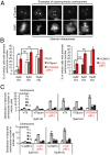
The impact of HR deficiency or very low HU doses (5 or 10 μM) on metaphases with aberrant centrosome number monitored with γ-tubulin antibody. (A) Examples of labeled centrosomes in mitotic cells (chromosomal DAPI staining). (Left) Normal centrosome number (= 2); columns 2–5, aberrant centrosomes number (unequal 2), causing metaphase alterations (see DNA labeling). (Scale bars: 10 μm.) (B) Frequency of mitotic cells with aberrant centrosome number. Left histograms, V79 cells and derivatives; right histograms, V-C8 cells and derivatives. The mean value ± SD was calculated from at least three independent experiments: *P < 0.05; **P < 0.01; ***P < 0.001; ns, not significant. In total, 150 mitoses were scored for each experiment and condition. (C) Centrosome number distribution in mitotic cells. Upper histograms, V79 cells and derivatives; lower histograms, V-C8 cells and derivatives. In total, 100–150 mitotic cells per cell line were analyzed per condition.
Fig. 3.
Fig. 3.
The impact of HR deficiency or 5 μM HU on mitosis duration. (A) Chromosome segregation kinetics. Example of time-lapse video microscopy of y-H2B-GFP–tagged wild-type (Top) and HR defective (Middle and Bottom) cells during a complete mitotic cycle. (Scale bars: 10 μm.) (B) Median kinetics of the different mitosis phase as measured by time-lapse video microscopy. For analysis, mitosis was clustered into prophase to metaphase and metaphase to anaphase. Left histogram, V79 cells and derivatives; right histogram, V-C8 cells and derivatives. *P < 0.05; **P < 0.01; ***P < 0.001; ns, not significant. At least 100 cells were analyzed per condition.
Fig. 4.
Fig. 4.
The impact of HR deficiency or very low HU doses (5 or 10 μM) on chromosome segregation. (A) Chromatin bridges. (A Upper) Example of anaphase chromatin bridge (see also Fig. 3A, Middle). (Scale bars: 10 μm.) (A Lower) Frequency of mitotic cells with chromatin bridges. Shown are V79 cells and derivatives (A Left) and V-C8 cells and derivatives (A Right). The mean value ± SD from three independent experiments was calculated. *P < 0.05; **P < 0.01; ***P < 0.001; ns, not significant. In total, 150 mitoses were scored per condition. (B) The frequency of aberrant mitosis in V79 cells and derivatives (Left) and V-C8 cells and derivatives (Right). *P < 0.05; **P < 0.01; ***P < 0.001; ns, not significant. The mean value ± SD of three independent experiments was calculated. In total, 150 mitoses were scored per condition.
Fig. 5.
Fig. 5.
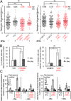
The effect of dNs addition on replication fork speed and the frequency of mitosis with aberrant centrosome number. (A) Replication fork speed distribution in V79 cells and derivatives (Left) and V-C8 cells and derivatives (Right) is presented. The numbers correspond to the median replication speed. Median and P values are indicated (*P < 0.05; **P < 0.01; ***P < 0.001). Median values are represented as horizontal black lines. Seventy to 145 fibers were scored per condition. (B) The quantification of mitotic cells with aberrant centrosome number (unequal 2) using γ-tubulin labeling. Left histogram, V79 cells and derivatives; right histogram, V-C8 cells and derivatives. The mean value ± SD of three independent experiments was calculated. *P < 0.05; **P < 0.01; ***P < 0.001; ns, not significant. In total, 150 mitoses were scored per condition. (C) Centrosome number distribution in mitotic cells monitored by immunofluorescence using γ-tubulin labeling. V79 cells and derivatives (Left); V-C8 cells and derivatives (Right). At least 100 mitotic cells were analyzed per condition.
Fig. 6.
Fig. 6.
RF deceleration alters global chromosome segregation. Different causes such as dNTP shortage, polymerase inhibition, and HR deficiency cause RF deceleration, which elicits the firing of RFs. At such low or endogenous stresses, cells do not arrest and, thus, progress through the G2 phase with incompletely replicated DNA. RF deceleration exacerbates these processes leading to chromatin bridges in nonreplicated regions. However, these cells are blocked at metaphase. Bypassing this arrest (in an “adaptation-like” process) causes abnormal mitosis including MEC, anaphase chromatin bridges and multipolar cells, which results in uneven chromosome segregation and aneuploidy in the whole genome, even for replicated chromosomes.

References

    1. Bartkova J, et al. DNA damage response as a candidate anti-cancer barrier in early human tumorigenesis. Nature. 2005;434(7035):864–870. - PubMed
    1. Deng G, Lu Y, Zlotnikov G, Thor AD, Smith HS. Loss of heterozygosity in normal tissue adjacent to breast carcinomas. Science. 1996;274(5295):2057–2059. - PubMed
    1. Gorgoulis VG, et al. Activation of the DNA damage checkpoint and genomic instability in human precancerous lesions. Nature. 2005;434(7035):907–913. - PubMed
    1. Krämer A. Centrosome aberrations—hen or egg in cancer initiation and progression? Leukemia. 2005;19(7):1142–1144. - PubMed
    1. Lakhani SR, et al. Genetic alterations in ‘normal’ luminal and myoepithelial cells of the breast. J Pathol. 1999;189(4):496–503. - PubMed

Publication types

LinkOut - more resources