. 2013:2013:510451.
doi: 10.1155/2013/510451. Epub 2013 Nov 21.

Poly (ADP-ribose) polymerase mediates diabetes-induced retinal neuropathy

Affiliations

Poly (ADP-ribose) polymerase mediates diabetes-induced retinal neuropathy

Ghulam Mohammad et al. Mediators Inflamm. 2013.

Abstract

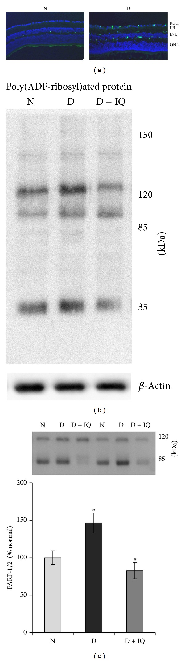
Retinal neuropathy is an early event in the development of diabetic retinopathy. One of the potential enzymes that are activated by oxidative stress in the diabetic retina is poly (ADP-ribose) polymerase (PARP). We investigated the effect of the PARP inhibitor 1,5-isoquinolinediol on the expression of the neurodegeneration mediators and markers in the retinas of diabetic rats. After two weeks of streptozotocin-induced diabetes, rats were treated with 1,5-isoquinolinediol (3 mg/kg/day). After 4 weeks of diabetes, the retinas were harvested and the levels of reactive oxygen species (ROS) were determined fluorometrically and the expressions of PARP, phosporylated-ERK1/2, BDNF, synaptophysin, glutamine synthetase (GS), and caspase-3 were determined by Western blot analysis. Retinal levels of ROS, PARP-1/2, phosphorylated ERK1/2, and cleaved caspase-3 were significantly increased, whereas the expressions of BDNF synaptophysin and GS were significantly decreased in the retinas of diabetic rats, compared to nondiabetic rats. Administration of 1,5-isoquinolinediol did not affect the metabolic status of the diabetic rats, but it significantly attenuated diabetes-induced upregulation of PARP, ROS, ERK1/2 phosphorylation, and cleaved caspase-3 and downregulation of BDNF, synaptophysin, and GS. These findings suggest a beneficial effect of the PARP inhibitor in increasing neurotrophic support and ameliorating early retinal neuropathy induced by diabetes.


Figures

Figure 1
Figure 1
Effect of 1,5-isoquinolinediol on retinal poly(ADP-ribose) polymerase (PARP) expression in diabetes. (a) Cryosections from normal (N) and diabetic (D) retina were subjected to immunostaining using anti-PARP antibodies (green) and DAPI (blue) was used to stain the nuclei. The sections were imaged at 20x magnification using Olympus BX-UCB fluorescent microscope. RGC: retinal ganglion cell; IPL: inner plexiform layer; INL: inner nuclear layer; ONL: outer nuclear layer. (b) Representative Western blot analyses of retinal poly(ADP-ribosyl)ated proteins in control and diabetic rats maintained with and without PARP inhibitor treatment. (c) PARP activation was measured by western blot technique, and the ratio of active PARP-2 (85 KDa) and pro-PARP-1 (120 kDa) was calculated in control and diabetic rats maintained with and without PARP inhibitor treatment. Measurements were made in duplicate in six to eight rats in each group. Western blots are representative of three different experiments. Results are expressed as mean ± SD. Values obtained from nondiabetic rats are considered as 100%. N: nondiabetic rat; D: diabetic rat; D + IQ: diabetic rat treated with 1,5-isoquinolinediol. *P < 0.05 compared with nondiabetic rat. # P < 0.05 compared with diabetic rat.
Figure 2
Figure 2
Effect of 1,5-isoquinolinediol on retinal ERK1/2 activation in diabetes. Relative abundance of phosphorylated ERK1/2 (p-ERK1/2) and total ERK1/2 (t-ERK1/2) was determined by Western blotting, followed by densitometry. Data are expressed as percentage change in phosphorylation over t-ERK1/2 and are expressed as mean ± SD. Values obtained from nondiabetic rats are considered as 100%. N: nondiabetic rat; D: diabetic rat; D + IQ: diabetic rat treated with 1,5-isoquinolinediol. *P < 0.05 compared with nondiabetic rat. # P < 0.05 compared with diabetic rat.
Figure 3
Figure 3
Effect of 1,5-isoquinolinediol on retinal brain-derived neurotrophic factor (BDNF) expression in diabetes. The expression of BDNF in retinal homogenate was determined by Western blotting technique. The histogram represents the mean band intensity (from 6–8 rats in each group) of BDNF adjusted to the intensity of β-actin in the same sample. Values obtained from nondiabetic rats are considered as 100%. N: nondiabetic rat; D: diabetic rat; D + IQ: diabetic rat treated with 1,5-isoquinolinediol. *P < 0.05 compared with nondiabetic rat. # P < 0.05 compared with diabetic rat.
Figure 4
Figure 4
Effect of 1,5-isoquinolinediol on retinal synaptophysin expression in diabetes. The expression of synaptophysin in retinal homogenates was determined by Western blotting technique. The histogram represents the mean band intensity (from 6–8 rats in each group) of synaptophysin adjusted to the intensity of β-actin in the same sample. Values obtained from nondiabetic rats are considered as 100%. N: nondiabetic rat; D: diabetic rat; D + IQ: diabetic rat treated with 1,5-isoquinolinediol. *P < 0.05 compared with nondiabetic rat. # P < 0.05 compared with diabetic rat.
Figure 5
Figure 5
Effect of 1,5-isoquinolinediol on retinal GS expression in diabetes. The expression of GS in retinal homogenates was determined by Western blotting technique. The histogram represents the mean band intensity (from 6–8 rats in each group) of GS adjusted to the intensity of β-actin in the same sample. Values obtained from nondiabetic rats are considered as 100%. N: nondiabetic rat; D: diabetic rat; D + IQ: diabetic rat treated with 1,5-isoquinolinediol. *P < 0.05 compared with nondiabetic rat. # P < 0.05 compared with diabetic rat.
Figure 6
Figure 6
Effect of 1,5-isoquinolinediol on retinal cleavage of caspase-3 in diabetes. Cleavage of caspase-3 was measured by Western blot analysis, and the ratio of active caspase-3 (17 KDa) and pro-caspase-3 (37 kDa) was calculated in control and diabetic rats maintained with and without PARP inhibitor treatment. Measurements were made in duplicate in six to eight rats in each group. Western blots are representative of three different experiments. Results are expressed as mean ± SD. Values obtained from nondiabetic rats are considered as 100%. N: nondiabetic rat; D: diabetic rat; D + IQ: diabetic rat treated with 1,5-isoquinolinediol. *P < 0.05 compared with nondiabetic rat. # P < 0.05 compared with diabetic rat.
Figure 7
Figure 7
Effect of 1,5-isoquinolinediol on retinal reactive oxygen species (ROS) levels in diabetes. Freshly prepared retinal homogenates were incubated with DCHFDA (5 μM) for 30 min. An equal amount of protein was used to quantitate 2′,7′-dichlorofluorescein fluorescence. N: nondiabetic rat; D: diabetic rat; D + IQ: diabetic rat treated with 1,5-isoquinolinediol. Data are expressed as percent control and mean ± SD from retina from 5-6 rats in each group. *P < 0.05 compared with nondiabetic rat. # P < 0.05 compared with diabetic rat.
Figure 8
Figure 8
Diabetes-induced retinal PARP-1/2 activation were prevented by apocynin. PARP-1/2 expression in retina was quantified by Western blotting analysis using β-actin as a loading protein. Each measurement was made in duplicate or triplicate. The values are represented as mean ± SD of 5–7 rats in each of the three groups. N: normal, D: Diabetes, D + Apo: Diabetic rat received apocynin. *P < 0.05 compared to normal and # P < 0.05 compared to diabetes.

Similar articles

Cited by

References

    1. Seki M, Tanaka T, Nawa H, et al. Involvement of brain-derived neurotrophic factor in early retinal neuropathy of streptozotocin-induced diabetes in rats: therapeutic potential of brain-derived neurotrophic factor for dopaminergic amacrine cells. Diabetes. 2004;53(9):2412–2419. - PubMed
    1. Barber AJ. A new view of diabetic retinopathy: a neurodegenerative disease of the eye. Progress in Neuro-Psychopharmacology and Biological Psychiatry. 2003;27(2):283–290. - PubMed
    1. Sasaki M, Ozawa Y, Kurihara T, et al. Neurodegenerative influence of oxidative stress in the retina of a murine model of diabetes. Diabetologia. 2010;53(5):971–979. - PMC - PubMed
    1. Simó R, Hernández C, European consortium for the early treatment of diabetic retinopathy (EUROCONDOR) Neurodegeneration is an early event in diabetic retinopathy: therapeutic implications. British Journal of Ophthalmology. 2012;96(10):1285–1290. - PubMed
    1. Santos JM, Mohammad G, Zhong Q, Kowluru RA. Diabetic retinopathy, superoxide damage and antioxidants. Current Pharmaceutical Biotechnology. 2011;12(3):352–361. - PMC - PubMed

MeSH terms