

Herlyn-Werner-Wunderlich syndrome presenting with infertility: Role of MRI in diagnosis
- PMID: 24347855
- PMCID: PMC3843333
- DOI: 10.4103/0971-3026.120283
Herlyn-Werner-Wunderlich syndrome presenting with infertility: Role of MRI in diagnosis
Abstract
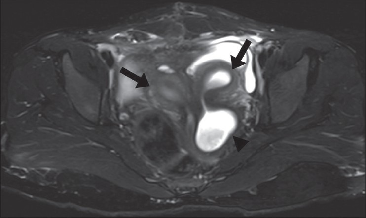
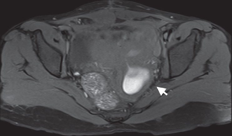
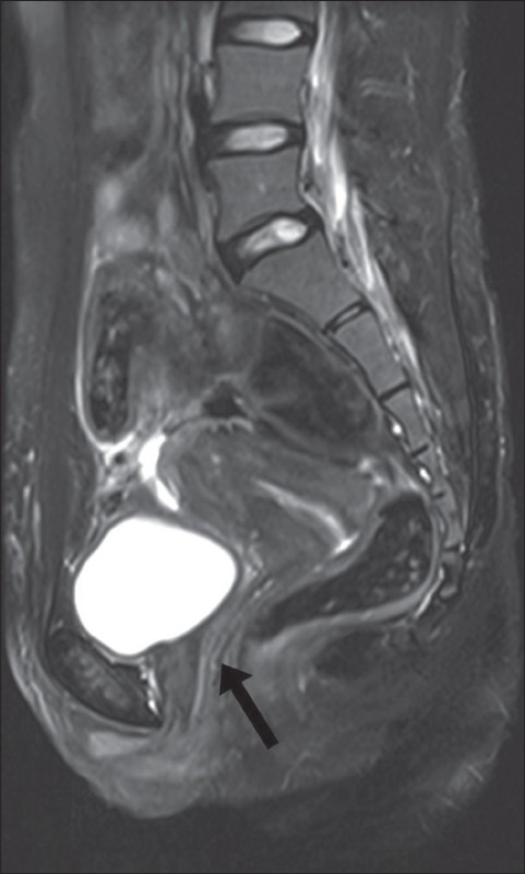
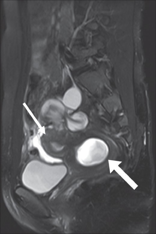
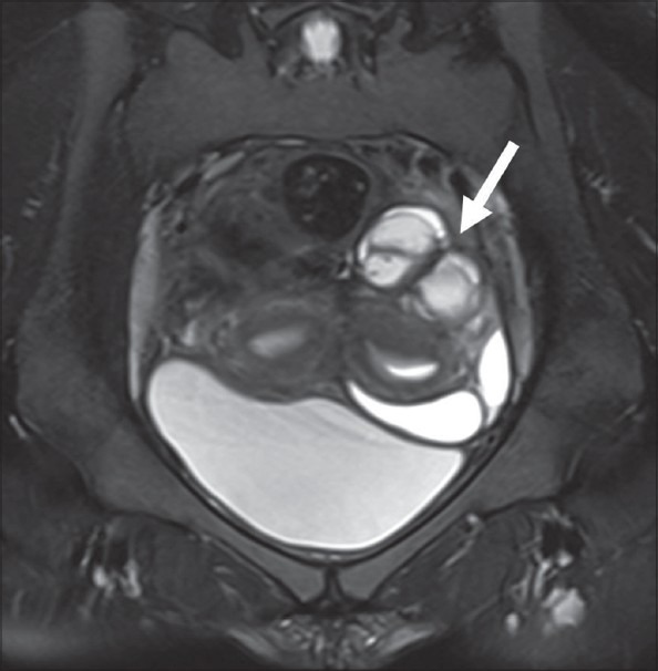
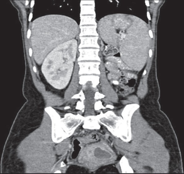
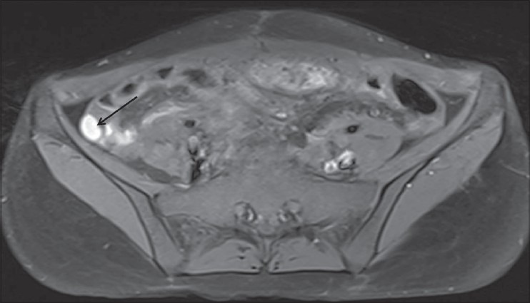
Herlyn-Werner-Wunderlich syndrome (HWWS), characterized by uterus didelphys, obstructed hemivagina, and ipsilateral renal agenesis, is an uncommon combined Mullerian and mesonephric duct anomaly, and its presentation in adulthood is even rarer. We report here a 22-year-old female presenting with primary infertility where magnetic resonance imaging (MRI) suggested the diagnosis of HWWS with endometriosis. In a patient of infertility with endometriosis and unilateral renal agenesis, diagnosis of HWWS should be suspected and MRI is the investigation of choice for such anomalies.
Keywords: Endometriosis; Herlyn-Werner-Wunderlich syndrome; infertility; magnetic resonance imaging; mullerian anomaly.
Conflict of interest statement
Figures
References
-
- Wu TH, Wu TT, Ng YY, Ng SC, Su PH, Chen JY, et al. Herlyn-Werner-Wunderlich Syndrome consisting of uterine didelphys, obstructed hemivagina and ipsilateral renal agenesis in a newborn. Pediatr Neonatol. 2012;53:68–71. - PubMed
-
- Jeong JH, Kim YJ, Chang CH, Choi HI. A case of Herlyn-Werner-Wunderlich syndrome with recurrent hematopyometra. J Womens’ Med. 2009;2:77–9.
-
- Asha B, Manila K. An unusual presentation of uterus didelphys with obstructed hemivagina with ipsilateral renal agenesis. Fertil Streril. 2008;849:e9–10. - PubMed
-
- Mandava A, Prabhakar RR, Smitha S. OHVIRA Syndrome (obstructed hemivagina and ipsilateral renal anomaly) with Uterus Didelphys, an Unusual Presentation. J Pediatr Adolesc Gynecol. 2012;25:e23–5. - PubMed
Publication types
LinkOut - more resources
Full Text Sources
Other Literature Sources