

Rare complications of cesarean scar
- PMID: 24347858
- PMCID: PMC3843336
- DOI: 10.4103/0971-3026.120265
Rare complications of cesarean scar
Abstract
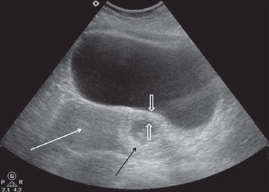
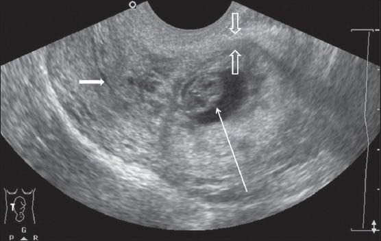
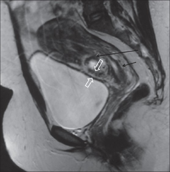
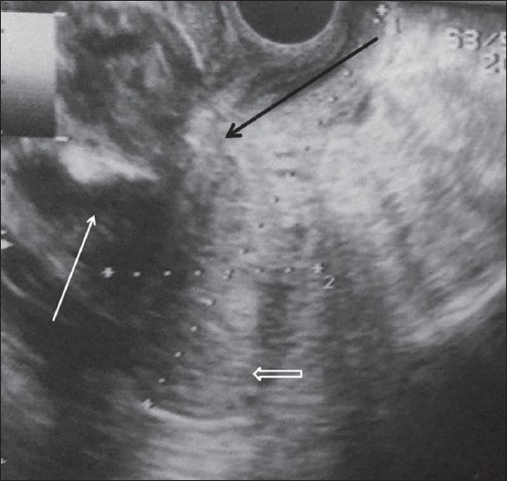
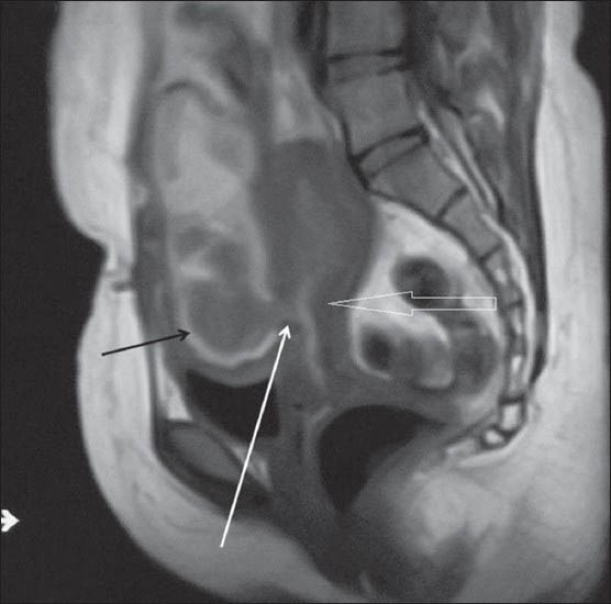
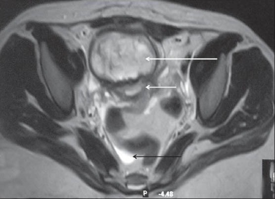
Cesarean scar pregnancy (CSP) and cesarean scar dehiscence (CSD) are the most dreaded complications of cesarean scar (CS). As the incidence of CS is increasing worldwide, so is the incidence of CSP, especially in cases with assisted reproduction techniques. It is of utmost importance to diagnose CSP in the early first trimester, as it can lead to myometrial rupture with fatal outcome. On the other hand, CSD may be encountered during pregnancy or in the postpartum period. CSD in the postpartum period is very rare and can cause secondary postpartum hemorrhage (PPH) leading to increased maternal morbidity or even death if not diagnosed and managed promptly. Both complications can be diagnosed on ultrasonography (USG) and confirmed on magnetic resonance imaging (MRI). These two conditions carry high morbidity and mortality. In this article, we highlight the role of imaging in the early diagnosis and management of these conditions.
Keywords: Cesarean scar dehiscence; cesarean scar pregnancy; magnetic resonance imaging; ultrasonography.
Conflict of interest statement
Figures
Similar articles
-
A novel method for typing of cesarean scar pregnancy based on size of cesarean scar diverticulum and its significance in clinical decision-making.J Obstet Gynaecol Res. 2020 May;46(5):707-714. doi: 10.1111/jog.14226. Epub 2020 Mar 9. J Obstet Gynaecol Res. 2020. PMID: 32153107
-
Magnetic resonance imaging as an adjunct to ultrasound in evaluating cesarean scar ectopic pregnancy.J Clin Imaging Sci. 2013 Mar 29;3:16. doi: 10.4103/2156-7514.109758. Print 2013. J Clin Imaging Sci. 2013. PMID: 23814688 Free PMC article.
-
Comparison of ultrasonic diagnosis of cesarean scar defects at different timepoints following cesarean section.Quant Imaging Med Surg. 2024 Aug 1;14(8):5490-5498. doi: 10.21037/qims-24-531. Epub 2024 Jul 26. Quant Imaging Med Surg. 2024. PMID: 39144005 Free PMC article.
-
Pitfalls in Ultrasound Diagnosis of Cesarean Scar Pregnancy.J Obstet Gynaecol India. 2018 Jun;68(3):164-172. doi: 10.1007/s13224-016-0956-1. Epub 2017 Jan 12. J Obstet Gynaecol India. 2018. PMID: 29895994 Free PMC article. Review.
-
Application of ultrasonography in the diagnosis and treatment of cesarean scar pregnancy.Clin Chim Acta. 2018 Nov;486:291-297. doi: 10.1016/j.cca.2018.08.012. Epub 2018 Aug 11. Clin Chim Acta. 2018. PMID: 30102898 Review.
Cited by
-
Post-caesarean Haematomas, Septic Collections and Wound Disruptions- Re-Laparotomy Based on Abdominal Imaging.J Clin Diagn Res. 2016 Nov;10(11):QJ01-QJ02. doi: 10.7860/JCDR/2016/20920.8833. Epub 2016 Nov 1. J Clin Diagn Res. 2016. PMID: 28050453 Free PMC article. No abstract available.
-
Use of Magnetic Resonance Imaging (MRI) in the Management of Diagnostic Uncertainty in Low-Resource Settings: A Case Report of Cesarean Ectopic Pregnancy in a Tertiary Hospital in Ghana.Am J Case Rep. 2020 Dec 28;21:e927496. doi: 10.12659/AJCR.927496. Am J Case Rep. 2020. PMID: 33370250 Free PMC article.
References
-
- Larsen JV, Janowski K, Krolilowski A. Secondary post partum haemorrhage due to uterine wound dehiscence. Cent Afr J Med. 1995;41:294–6. - PubMed
-
- Seow KM, Huang LW, Lin YH, Yan-Sheng Lin M, Tsai YL, Hwang JL. Cesarean scar pregnancy: Issues in management. Ultrasound Obstet Gynaecol. 2004;23:247–53. - PubMed
-
- Khalifa Y, Redgment CJ, Yazdani N, Taranissi M, Craft IL. Pregnancy. Intramural pregnancy following difficult embryo transfer. Hum Reprod. 1994;9:2427–8. - PubMed
-
- Maldjian C, Milestone B, Schnall M, Smith R. MR appearance of uterine dehiscence in the post-cesarean section patient. J Comput Assist Tomogr. 1998;22:738–41. - PubMed
Publication types
LinkOut - more resources
Full Text Sources
Other Literature Sources
Research Materials