

Ocular cysticercosis in a 32-year-old man in Abuja: ultrasonic features as an aid in diagnosis
- PMID: 24348016
- PMCID: PMC3857263
- DOI: 10.2147/OPTH.S52690
Ocular cysticercosis in a 32-year-old man in Abuja: ultrasonic features as an aid in diagnosis
Abstract
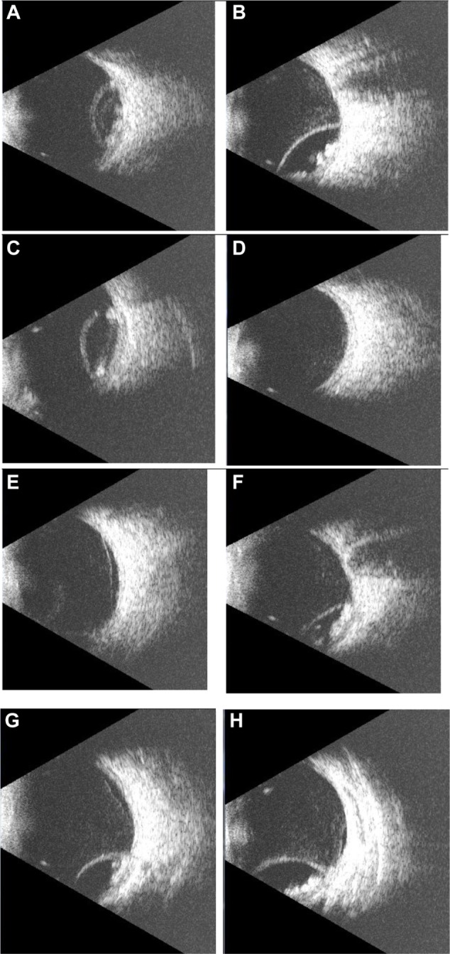
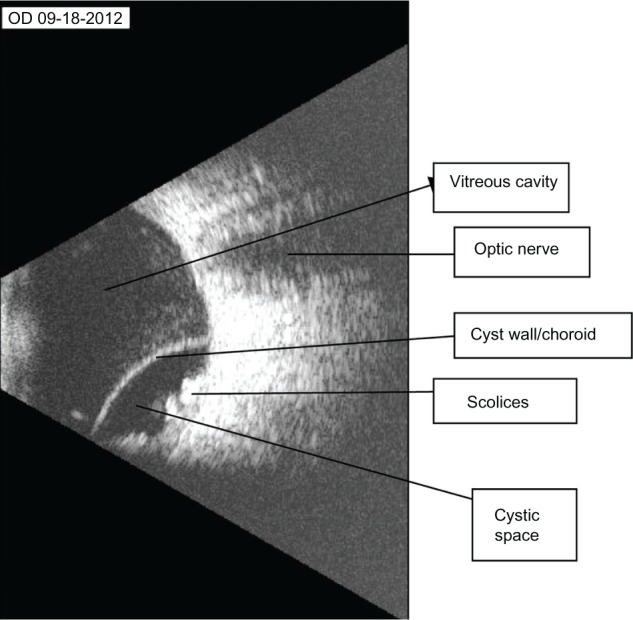
We report the case of a 32-year-old man suffering from intraocular cysticercosis, with special emphasis on the use of B-scan ultrasound in the diagnosis and management of the condition. An 8000 B-Scan Scanmate was used to obtain the ultrasound images. The patient had worked on a pig farm a few years before presentation. He presented with shadows seen in the right eye. Binocular indirect ophthalmoscopy revealed that he had a choroidal detachment in the right eye inferotemporally. B-scan ultrasound revealed a subretinal subchoroidal cyst with a thick wall containing well defined intracystic echogenic entities representing scolices, and an associated retinal detachment. These findings appear to be pathognomonic. Excision of the cyst through a trans-scleral approach revealed a yellowish serous fluid, with scolices of cysticercus later confirmed histologically. B-scan ultrasound is extremely useful in the diagnosis of ocular cysticercosis and the findings can be pathognomonic.
Keywords: B-scan ultrasound; Nigeria; ocular cysticercosis.
Figures
References
-
- Weka RP, Ikeh EI. Seroprevalence of cysticercosis and intestinal parasitism in pigs in Jos Metropolis. J Anim Vet Adv. 2009;8:883–887.
-
- Centers for Disease Control and Prevention Recommendations of International Task Force for Disease Eradication (ITFDE) MMWR Recomm Rep. 1993;42:1–38. - PubMed
-
- Hoberg EP. Taenia tapeworms: their biology, evolution and socioeconomic significance. Microbes Infect. 2002;4:859–866. - PubMed
-
- Centers for Disease Control and Prevention The life cycle of Taenia solium. [Accessed September 22, 2013]. Available from: http://www.dpd.cdc.gov/dpdx/html/cysticercosis.htm.
Publication types
LinkOut - more resources
Full Text Sources
Other Literature Sources
