Review
. 2013 Nov 20:2013:683685.
doi: 10.1155/2013/683685.

Ultrasound for the anesthesiologists: present and future

Affiliations
Review

Ultrasound for the anesthesiologists: present and future

Abdullah S Terkawi et al. ScientificWorldJournal. .

Abstract

Ultrasound is a safe, portable, relatively inexpensive, and easily accessible imaging modality, making it a useful diagnostic and monitoring tool in medicine. Anesthesiologists encounter a variety of emergent situations and may benefit from the application of such a rapid and accurate diagnostic tool in their routine practice. This paper reviews current and potential applications of ultrasound in anesthesiology in order to encourage anesthesiologists to learn and use this useful tool as an adjunct to physical examination. Ultrasound-guided peripheral nerve blockade and vascular access represent the most popular ultrasound applications in anesthesiology. Ultrasound has recently started to substitute for CT scans and fluoroscopy in many pain treatment procedures. Although the application of airway ultrasound is still limited, it has a promising future. Lung ultrasound is a well-established field in point-of-care medicine, and it could have a great impact if utilized in our ORs, as it may help in rapid and accurate diagnosis in many emergent situations. Optic nerve sheath diameter (ONSD) measurement and transcranial color coded duplex (TCCD) are relatively new neuroimaging modalities, which assess intracranial pressure and cerebral blood flow. Gastric ultrasound can be used for assessment of gastric content and diagnosis of full stomach. Focused transthoracic (TTE) and transesophageal (TEE) echocardiography facilitate the assessment of left and right ventricular function, cardiac valve abnormalities, and volume status as well as guiding cardiac resuscitation. Thus, there are multiple potential areas where ultrasound can play a significant role in guiding otherwise blind and invasive interventions, diagnosing critical conditions, and assessing for possible anatomic variations that may lead to plan modification. We suggest that ultrasound training should be part of any anesthesiology training program curriculum.


Figures

Figure 1
Figure 1
Concept of ultrasonography: *tissues that allow the beam to pass easily (e.g., containing fluids or blood) create only little echo (hypoechoic) and appear black on the screen, while tissues that allows less beam to pass (e.g., fat and bone) create stronger echoes (hyperechoic) and thus appear white on the screen; **linear transducers have a higher frequency (10–15 MHz) and are usually used for superficial structures; curved transducers have a lower frequency (4–8 MHz) and are usually used for deeper structures.
Figure 2
Figure 2
Vocal cords assessment: SM: strap muscles; TC: thyroid cartilage; AC: anterior commissure; PC: posterior commissure; and VC: vocal cords. (a) Vocal cords are abducted on inspiration, (b) adducted partially during expiration, (c) and are tightly closed when asking the patient to say “Eeeee.” Linear transducer was placed transversely on the midline of the cricothyroid membrane.
Figure 3
Figure 3
Normal lung ultrasound: (a) 2D “red arrows” point to the pleura, where the normal “sliding sign” should be seen, while the “yellow arrows” represent the A-lines that are normal reverberation from the pleura. (b) M-mode shows the “seashore sign.” Pneumothorax: (c) 2D; absence of lung sliding, (d) M-mode; “stratosphere sign” or “barcode sign,” lung point may also be seen during inspiration and represents the border between pneumothorax and normal pleura. Cardiac pulmonary edema: (e) homogeneous distribution of B-lines (yellow arrows), normal sliding, and no spared areas. Acute respiratory distress syndrome (ARDS): (f) “patchy” distribution of B-lines, reduced/abolished sliding, spared areas, and peripheral consolidations.
Figure 4
Figure 4
Pulmonary embolism: ((a) lung ultrasound) peripheral, triangular, and pleural based hypoechoic lesions (yellow arrows); ((b) transthoracic echo, apical view) it shows right ventricular (RV) dilation, RV hypokinesia, septal flattening, and tricuspid regurgitation. IVS: interventricular septum; TV: tricuspid valve; LV: left ventricle; RA: right atrium; LA: left atrium.
Figure 5
Figure 5
Optic nerve sheath: (a) normal diameter and (b) large diameter that represents increase intracranial pressure. ONSD: optic nerve sheath diameter. It is usually measured 3 mm behind the retina. Ultrasound pupillary light reflex: (c) diameter of the pupil before shining light to the contralateral eye and (d) the pupil constricted after shining the light.
Figure 6
Figure 6
Transcranial color coded duplex (TCCD): (a) middle cerebral artery (MCA) color Doppler and (b) MCA pulsed wave Doppler. S: systole, D: diastole.
Figure 7
Figure 7
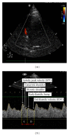
Basic transthoracic echo views: (a) left parasternal long axis, (b) apical, (c) subcostal, and (d) left parasternal short axis; aortic valve “Mercedes sign,” mitral valve “fish mouth sign,” and papillary muscles (two arrows), respectively, from left to right. RV: right ventricle, LV: left ventricle, LVOT: left ventricular outlet, RA: right atrium, LA: left atrium, AV: aortic valve, MV: mitral valve, and TV: tricuspid valve.

References

    1. Kline JP. Ultrasound guidance in anesthesia. AANA Journal. 2011;79(3):209–217. - PubMed
    1. Falyar CR. Ultrasound in anesthesia: applying scientific principles to clinical practice. AANA Journal. 2010;78(4):332–340. - PubMed
    1. Hatfield A, Bodenham A. Ultrasound: an emerging role in anaesthesia and intensive care. British Journal of Anaesthesia. 1999;83(5):789–800. - PubMed
    1. Kristensen MS. Ultrasonography in the management of the airway. Acta Anaesthesiologica Scandinavica. 2011;55(10):1155–1173. - PubMed
    1. Stefanidis K, Dimopoulos S, Nanas S. Basic principles and current applications of lung ultrasonography in the intensive care unit. Respirology. 2011;16(2):249–256. - PubMed

LinkOut - more resources