. 2013 Nov 11:2013:740420.
doi: 10.1155/2013/740420. eCollection 2013.

Islet-1 immunoreactivity in the developing retina of Xenopus laevis

Affiliations

Islet-1 immunoreactivity in the developing retina of Xenopus laevis

Guadalupe Álvarez-Hernán et al. ScientificWorldJournal. .

Abstract

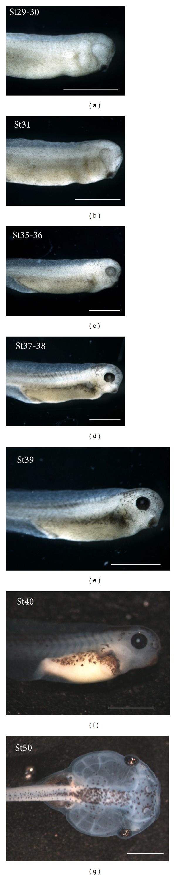
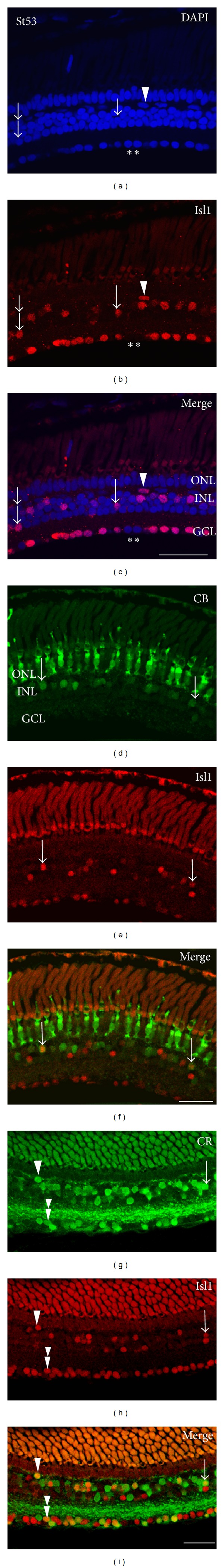
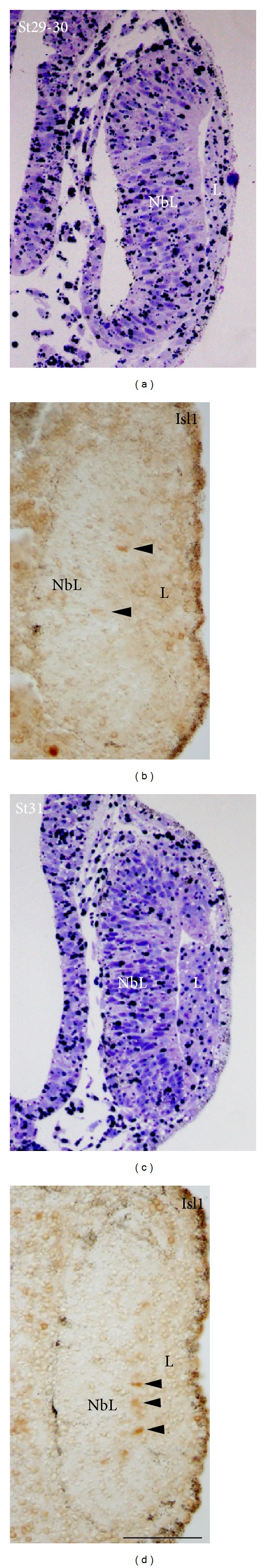
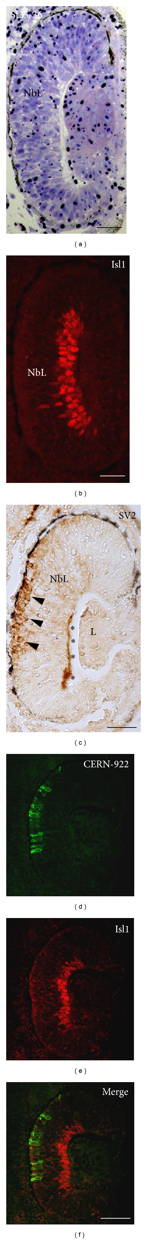
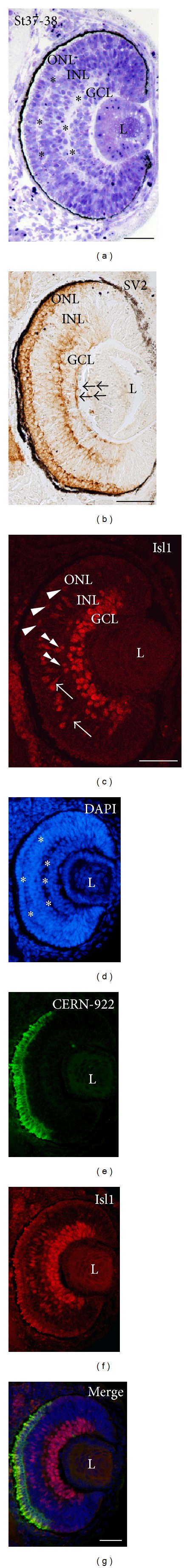
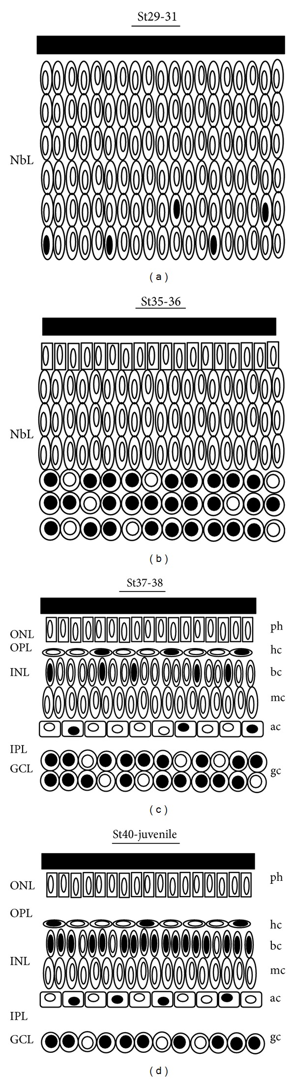
The LIM-homeodomain transcription factor Islet1 (Isl1) has been widely used as a marker of neuronal differentiation in the developing visual system of different classes of vertebrates, including mammals, birds, reptiles, and fish. In the present study, we analyzed the spatial and temporal distribution of Isl1-immunoreactive cells during Xenopus laevis retinal development and its relation to the formation of the retinal layers, and in combination with different markers of cell differentiation. The earliest Isl1 expression appeared at St29-30 in the cell nuclei of sparse differentiating neuroblasts located in the vitreal surface of the undifferentiated retina. At St35-36, abundant Isl1-positive cells accumulated at the vitreal surface of the neuroepithelium. As development proceeded and through the postmetamorphic juveniles, Isl1 expression was identified in subpopulations of ganglion cells and in subsets of amacrine, bipolar, and horizontal cells. These data together suggest a possible role for Isl1 in the early differentiation and maintenance of different retinal cell types, and Isl1 can serve as a specific molecular marker for the study of retinal cell specification in X. laevis.


Figures

Figure 1
Figure 1
Stereomicroscope images of selected lateral views of Xenopus laevis tailbud embryos ((a)–(f)) and a dorsal view of a free-swimming tadpole (g) according to developmental stages (St) of Nieuwkoop and Faber [37]. Scale bars: 1 mm.
Figure 2
Figure 2
Morphological features and expression patterns of Isl1 and other cell differentiation markers in the St53 Xenopus laevis central retina. (a)–(c) DAPI fluorescence combined with Isl1 immunofluorescence. DAPI staining showed well-organized retinal layers. Isl1 expressing cells in the INL could be characterized morphologically and topographically as horizontal (arrowheads), bipolar (arrows), or amacrine cells (double arrows). Notice that not all cells located in the GCL expressed Isl1 (asterisks). (d)–(f) A few Isl1-positive bipolar cells also expressed CB (arrows). (g)–(i) Subpopulations of Isl1-positive horizontal (arrowheads), bipolar (arrows), and ganglion cells (double arrowheads) also expressed CR. GCL: ganglion cell layer; INL: inner nuclear layer; ONL: outer nuclear layer. Scale bars: 100 μm.
Figure 3
Figure 3
Morphological features and expression patterns of Isl1 in the St29/30 ((a), (b)) and St31 ((c), that Isl1 is also expressed by undi (d)) Xenopus laevis retina. ((a), (c)) Toluidine blue-stained transverse retinal resin sections showed that the neural retina had the structure of pseudostratified columnar epithelium. Notice that during these early stages abundant dark-stained granules were observed in the cytoplasm of the neuroepithelial cells. (b),(d) Isl1 immunoreactivity was present in sparse nuclei mainly located near the vitreal surface (arrowheads). L: lens; NbL: neuroblastic layer. Scale bar: 50 μm.
Figure 4
Figure 4
Morphological features and expression patterns of Isl1 and other cell differentiation markers in the St35/36 Xenopus laevis retina. (a) Toluidine blue-stained transverse retinal resin sections showed that neural retina remained undifferentiated during this stage. (b) Although no plexiform layers were observed, many Isl1-positive nuclei were mainly located near the inner surface of the neuroretina. (c) Immunoreactive ganglion cell axons for SV2 were observed in the vitreal surface of the retina (asterisks). Furthermore, immunoreactivity was also detected near the scleral surface of the undifferentiated retina (arrowheads). (d)–(f) CERN-922 antibody revealed that sparse photoreceptors were differentiating by this stage in the scleral surface of the central nonlayered retina ((d), (F)). L: lens; NbL: neuroblastic layer. Scale bars: 50 μm.
Figure 5
Figure 5
Morphological features and expression patterns of Isl1 and other cell differentiation markers in the St37/38 Xenopus laevis retina. (a) Toluidine blue-stained transverse retinal resin sections showed that the plexiform layers were observed across the central to mid-peripheral extent of the retina (asterisks). An immature GCL, 2-3 cells in depth, was also observed at this stage. (b) SV2 immunoproducts were observed in the OFL (double arrows), IPL, and ONL. (c) Abundant nuclei were immunoreactive for Isl1 in the GCL, but also in the INL. Thus, nuclei located in the amacrine cell layer (double arrowheads), bipolar cell layer (arrows), and horizontal cell layer (arrowheads) were detected with this antibody. (d)–(g) Incipient plexiform layers were also clearly distinguishable with the DAPI nucleic acid stain (asterisks in (d)). CERN-922 immunoreactivity paralleled the expression of Isl1 and extended towards the more mid-peripheral region of the retina. GCL: ganglion cell layer; INL: inner nuclear layer; L: lens; ONL: outer nuclear layer. Scale bars: 50 μm.
Figure 6
Figure 6
Morphological features and expression patterns of Isl1 and other cell differentiation markers in the St40 Xenopus laevis retina. (a) Toluidine blue-stained transverse retinal resin sections revealed that the typical multilayered structure was clearly distinguishable, showing morphological features of maturation. The CMZ was visible in the peripheralmost region. (b) Isl1 immunoreactivity was clearly established in abundant nuclei located in the GCL, whereas the INL contained Isl1 labeling that corresponded to horizontal (arrowhead), bipolar (arrow), and amacrine cells (double arrowhead). (c) SV2 expression in the plexiform layers extended to more peripheral regions. (d)–(f) The CERN-922 labeling in the ONL coursed in parallel with Isl1 immunoreactivity and reached the periphery by this stage. CMZ: ciliary marginal zone; GCL: ganglion cell layer; INL: inner nuclear layer; L: lens; ONH: optic nerve head; ONL: outer nuclear layer. Scale bars: 100 μm.
Figure 7
Figure 7
Schematic drawings of the Isl1 positive cells in the developing retina of Xenopus laevis. Black nuclei represent Isl1-immunostained cells. (a) Isl1 immunoreactivity during St29–31 was restricted to nuclei located near the vitreal surface of the NbL. (b) Although the retinal lamination was absent in the St35-36, cell differentiation was detected immunohistochemically. Abundant Isl1-positive cells were located in the vitreal half of the NbL. (c) At St37-38, retinal layering was completed in the central retina with the emergence of the plexiform layers. Isl1 immunoreactivity was detected in the GCL and in cell subpopulations located in the INL. (d) From St40 onwards Isl1 immunoreactivity was confined to subpopulations of ganglion, amacrine, bipolar, and horizontal cells. Ac: amacrine cell; bc: bipolar cell; gc: ganglion cell; GCL: ganglion cell layer; hc: horizontal cell; INL: inner nuclear layer; IPL: inner plexiform layer; L: lens; mc: Müller cell; NbL: neuroblastic layer; ONL: outer nuclear layer; OPL: outer plexiform layer; ph: photoreceptor.

Similar articles

Cited by

References

    1. Young RW. Cell proliferation during postnatal development of the retina in the mouse. Brain Research. 1985;353(2):229–239. - PubMed
    1. Cepko CL. The patterning and onset of opsin expression in vertebrate retinae. Current Opinion in Neurobiology. 1996;6(4):542–546. - PubMed
    1. Marquardt T, Ashery-Padan R, Andrejewski N, Scardigli R, Guillemot F, Gruss P. Pax6 is required for the multipotent state of retinal progenitor cells. Cell. 2001;105(1):43–55. - PubMed
    1. Wang SW, Kim BS, Ding K, et al. Requirement for math5 in the development of retinal ganglion cells. Genes and Development. 2001;15(1):24–29. - PMC - PubMed
    1. Cheng CW, Chow RL, Lebel M, et al. The Iroquois homeobox gene, Irx5, is required for retinal cone bipolar cell development. Developmental Biology. 2005;287(1):48–60. - PubMed

Publication types

MeSH terms

Substances

LinkOut - more resources