. 2013;9(12):e1003774.
doi: 10.1371/journal.ppat.1003774. Epub 2013 Dec 5.

Tmprss2 is essential for influenza H1N1 virus pathogenesis in mice

Affiliations

Tmprss2 is essential for influenza H1N1 virus pathogenesis in mice

Bastian Hatesuer et al. PLoS Pathog. 2013.

Erratum in

  • PLoS Pathog. 2014 Sep;10(9):e1004435. Pöhlman, Stefan [corrected to Pöhlmann, Stefan]

Abstract

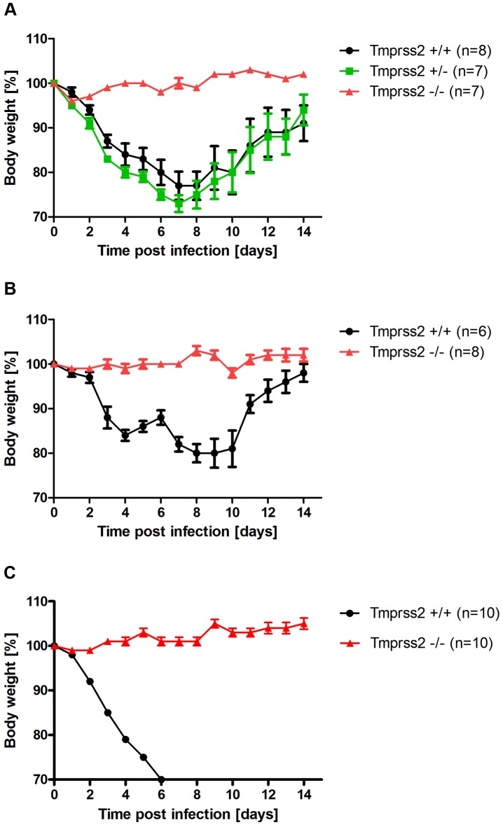
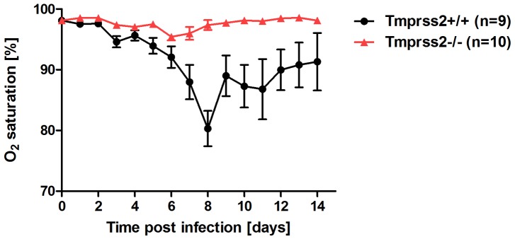
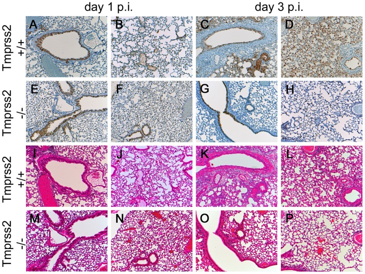
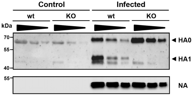
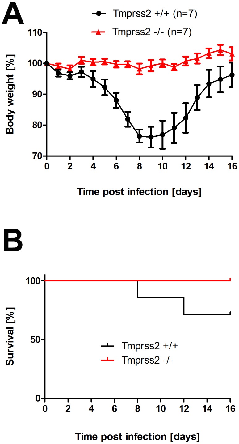
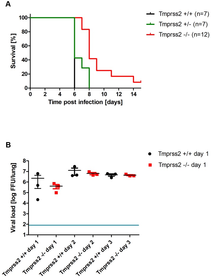
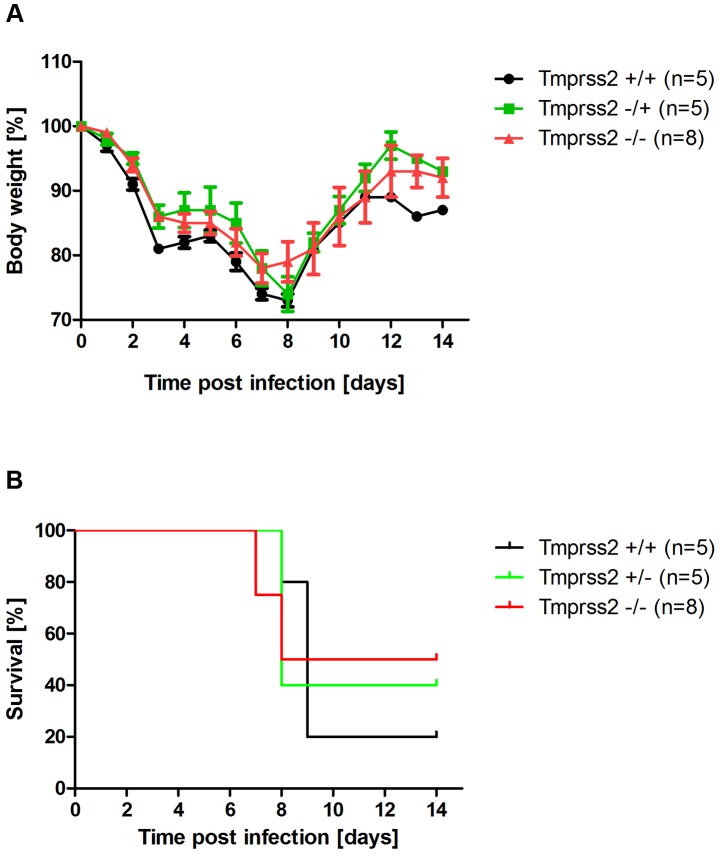
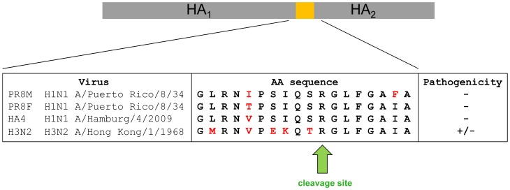
Annual influenza epidemics and occasional pandemics pose a severe threat to human health. Host cell factors required for viral spread but not for cellular survival are attractive targets for novel approaches to antiviral intervention. The cleavage activation of the influenza virus hemagglutinin (HA) by host cell proteases is essential for viral infectivity. However, it is unknown which proteases activate influenza viruses in mammals. Several candidates have been identified in cell culture studies, leading to the concept that influenza viruses can employ multiple enzymes to ensure their cleavage activation in the host. Here, we show that deletion of a single HA-activating protease gene, Tmprss2, in mice inhibits spread of mono-basic H1N1 influenza viruses, including the pandemic 2009 swine influenza virus. Lung pathology was strongly reduced and mutant mice were protected from weight loss, death and impairment of lung function. Also, after infection with mono-basic H3N2 influenza A virus body weight loss and survival was less severe in Tmprss2 mutant compared to wild type mice. As expected, Tmprss2-deficient mice were not protected from viral spread and pathology after infection with multi-basic H7N7 influenza A virus. In conclusion, these results identify TMPRSS2 as a host cell factor essential for viral spread and pathogenesis of mono-basic H1N1 and H3N2 influenza A viruses.


Conflict of interest statement

The authors have declared that no competing interests exist.

Figures

Figure 1
Figure 1. Tmprss2 is essential for spread and pathogenesis of H1N1 influenza viruses.
Eight to eleven weeks old female mice were infected with 2×10⁵ FFU mouse-adapted PR8M (H1N1; A), 2×10⁵ FFU HA4 (pH1N1, B), 2×10³ FFU mouse-adapted PR8F (H1N1; C). Body weight loss was monitored until day 14 p.i. Mice with a weight loss of more than 30% of the starting bodyweight were euthanized and recorded as dead. Weight loss data represent mean values +/− SEM. Note that only data of surviving mice are presented (e.g. about 50% of infected mice died after infection with PR8M, see Fig. S1). Body weight loss was significantly different between wild type and homozygous mutant mice at day 6 p.i. (p<0.0001 for PR8M infected mice, and p<0.0001 for HA4 infected mice, using the non-parametric Mann Whitney U test) and between heterozygous and homozygous mutant mice (PR8M infected mice, p<0.0001 using the Mann Whitney U test).
Figure 2
Figure 2. Tmprss2−/− mutant mice do not exhibit drop in blood oxygen saturation.
Tmprss2−/− and wild type mice were infected with 2×10⁵ FFU PR8M virus and oxygen saturation in the peripheral blood was measured until day 14 p.i. Tmprss2−/− mice showed only a very mild drop in oxygen saturation whereas wild type mice exhibited a significant decrease that peaked at day 8 p.i.
Figure 3
Figure 3. Mild lung pathology and reduced viral spread is observed in Tmprss2−/− mutant mice.
Eight to twelve weeks old mice were infected intra-nasally with 2×10⁵ FFU of PR8M virus. Serial lung sections were stained at day 1 and day 3 p.i. with anti-influenza antibody and haematoxylin (A–H) or with haematoxylin/eosin (I–P). The overall lung tissues were more densely consolidated with larger numbers of infiltrating immune cells in wild type (I–L) compared to Tmprss2−/− mice (M–P). In addition, the airways of Tmprss2+/+ mice were surrounded by higher numbers neutrophils and macrophages (I–L) whereas airways of Tmprss2−/− mice showed lower numbers of immune cell infiltrations (M–P). Virus-infected cells at day 1 p.i. were observed mainly in bronchiolar regions in the lungs of both mice (A, B, E, F). Both wild type and mutant mice showed influenza-positive cells at day 3 p.i. (C, G). However, the overall number of infected cells was lower in Tmprss2−/− compared to wild type mice. Furthermore, infected cells were mostly limited to bronchiolar regions in Tmprss2−/− whereas in wild type mice the virus also spread significantly into the alveolar regions (D, H).
Figure 4
Figure 4. Viral load in the lungs of Tmprss2−/− mice after infection with H1N1 (PR8M) influenza A virus.
Eight to eleven weeks old female mice were infected with 2×10⁵ FFU of the PR8M virus. Infectious virus particles were determined in lung homogenates. Viral load was higher in infected wild type mice compared to infected homozygous mutant mice at days 1, 2 and 3 p.i. Individual values, mean and SEM are presented. Detection limit of the assay is at 80 infectious particles per lung indicated by the blue line. Day 1 p.i. n = 9 for Tmprss2−/−, n = 11 for Tmprss2+/+, day 2 p.i. n = 6 for Tmprss2−/−, n = 5 for Tmprss2+/+, day 3 p.i. n = 7 for Tmprss2−/−, n = 7 for Tmprss2+/+.
Figure 5
Figure 5. The hemagglutinin of H1N1 PR8M influenza virus is not processed in Tmprss2 knock-out mice.
BAL from infected wild type and Tmprss2−/− male mice was harvested at day 1 after infection with 2×10⁵ FFU PR8M and viral particles were concentrated by centrifugation through a sucrose cushion. As control, BAL from non-infected wild type and Tmprss2−/− was analyzed. Each sample was loaded undiluted (first lane), and in two dilutions (second lane 1∶1.33, third lane 1∶2). The virus-containing pellets were then analyzed for HA cleavage by Western blots. As loading control, the stripped membranes were incubated with anti-influenza A virus antibody confirming that equal amounts of proteins were loaded for respective undiluted and diluted samples.
Figure 6
Figure 6. Tmprss2 knock-out mice show reduced body weight loss and mortality after infection with low dose H3N2 influenza A virus infections.
Eight to eleven weeks old female mice were infected with 101 FFU mouse-adapted H3N2 influenza virus by intra-nasal application and bodyweight (A) and survival (B) was monitored until day 14 p.i. In addition to mice that were found dead, mice with a weight loss of more than 30% of the starting body weight were euthanized and recorded as dead. Homozygous Tmprss2 knock-out mice lost significantly less weight than wild type (e.g. p = 0,0006 at day 7, and p = 0,0006 at day 8, using MWU test) mice and showed reduced mortality compared to wild type mice, although this difference was not significant (using the log rank test).
Figure 7
Figure 7. Tmprss2 knock-out mice show reduced mortality after infection with high dose H3N2 influenza A virus infections.
Eight to eleven weeks old female mice were infected with 2×10³ FFU mouse-adapted H3N2 influenza virus by intra-nasal application and survival (A) was monitored until day 14 p.i. In addition to mice that were found dead, mice with a weight loss of more than 30% of the starting body weight were euthanized and recorded as dead. Infectious viral particles were determined in lung homogenates (B). Individual values, mean and SEM are presented. Detection limit of the assay is at 80 infectious particles per lung indicated by the blue line. Homozygous Tmprss2 knock-out mice showed significantly reduced mortality compared to wild type and heterozygote mice (p<0.0001 and p = 0.0032, respectively, using the log rank test). Viral load was not significantly different in infected wild type mice compared to infected homozygous mutant mice at days 1 to 3 p.i.
Figure 8
Figure 8. Murine Tmprss2 is not required for spread of H7N7 influenza A virus.
Eight to eleven weeks old female mice were infected with 2×10⁴ FFU mouse-adapted SC35M (H7N7) influenza virus by intra-nasal application and bodyweight (A) and survival (B) was monitored until day 14 p.i. In addition to mice that were found dead, mice with a weight loss of more than 30% of the starting bodyweight were euthanized and recorded as dead. No significant differences were observed in survival between heterozygous and homozygous mutant mice after H7N7 infections (using the log rank test).
Figure 9
Figure 9. Alignment of amino acid sequences of the protease loop region from H1N1 and H3N2 influenza A viruses.
Pathology is strongly reduced in Tmprss2−/− mice after infection with H1N1 virus and diminished after infection with H3N2 virus.

Similar articles

Cited by

References

    1. Hurt AC, Chotpitayasunondh T, Cox NJ, Daniels R, Fry AM, et al. (2012) Antiviral resistance during the 2009 influenza A H1N1 pandemic: public health, laboratory, and clinical perspectives. Lancet Infect Dis 12: 240–248. - PubMed
    1. Klenk HD, Rott R, Orlich M, Blodorn J (1975) Activation of influenza A viruses by trypsin treatment. Virology 68: 426–439. - PubMed
    1. Lazarowitz SG, Choppin PW (1975) Enhancement of the infectivity of influenza A and B viruses by proteolytic cleavage of the hemagglutinin polypeptide. Virology 68: 440–454. - PubMed
    1. Stieneke-Grober A, Vey M, Angliker H, Shaw E, Thomas G, et al. (1992) Influenza virus hemagglutinin with multibasic cleavage site is activated by furin, a subtilisin-like endoprotease. EMBO J 11: 2407–2414. - PMC - PubMed
    1. Kido H, Yokogoshi Y, Sakai K, Tashiro M, Kishino Y, et al. (1992) Isolation and characterization of a novel trypsin-like protease found in rat bronchiolar epithelial Clara cells. A possible activator of the viral fusion glycoprotein. J Biol Chem 267: 13573–13579. - PubMed

Publication types

Substances