. 2013;9(12):e1003816.
doi: 10.1371/journal.ppat.1003816. Epub 2013 Dec 12.

Phagocytosis escape by a Staphylococcus aureus protein that connects complement and coagulation proteins at the bacterial surface

Affiliations

Phagocytosis escape by a Staphylococcus aureus protein that connects complement and coagulation proteins at the bacterial surface

Ya-Ping Ko et al. PLoS Pathog. 2013.

Abstract

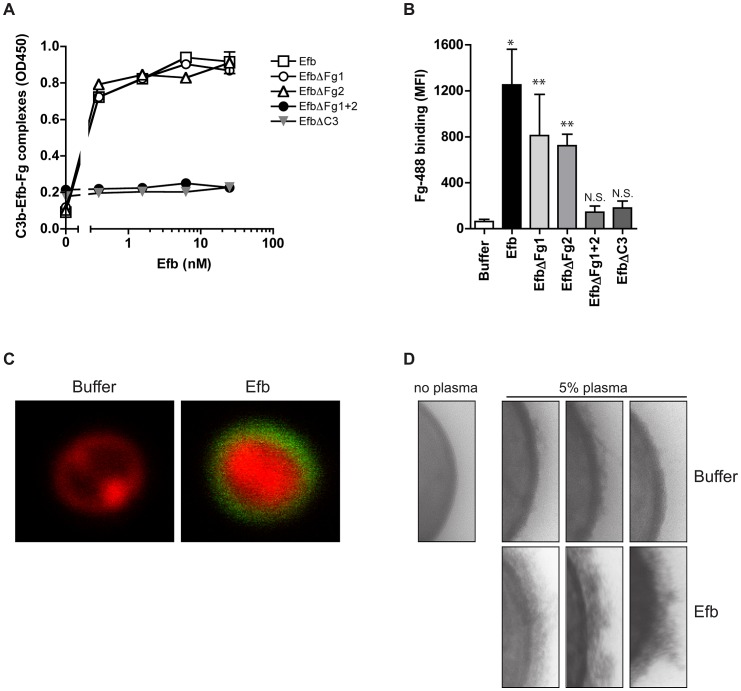
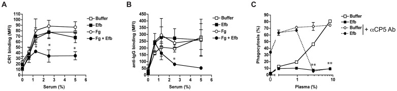
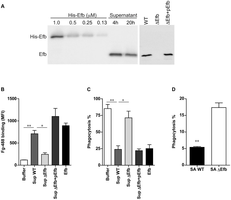
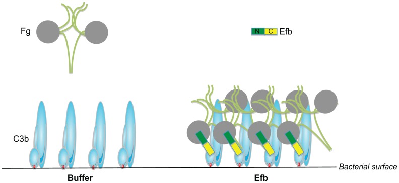
Upon contact with human plasma, bacteria are rapidly recognized by the complement system that labels their surface for uptake and clearance by phagocytic cells. Staphylococcus aureus secretes the 16 kD Extracellular fibrinogen binding protein (Efb) that binds two different plasma proteins using separate domains: the Efb N-terminus binds to fibrinogen, while the C-terminus binds complement C3. In this study, we show that Efb blocks phagocytosis of S. aureus by human neutrophils. In vitro, we demonstrate that Efb blocks phagocytosis in plasma and in human whole blood. Using a mouse peritonitis model we show that Efb effectively blocks phagocytosis in vivo, either as a purified protein or when produced endogenously by S. aureus. Mutational analysis revealed that Efb requires both its fibrinogen and complement binding residues for phagocytic escape. Using confocal and transmission electron microscopy we show that Efb attracts fibrinogen to the surface of complement-labeled S. aureus generating a 'capsule'-like shield. This thick layer of fibrinogen shields both surface-bound C3b and antibodies from recognition by phagocytic receptors. This information is critical for future vaccination attempts, since opsonizing antibodies may not function in the presence of Efb. Altogether we discover that Efb from S. aureus uniquely escapes phagocytosis by forming a bridge between a complement and coagulation protein.


Conflict of interest statement

The authors have declared that no competing interests exist.

Figures

Figure 1
Figure 1. Full-length Efb inhibits phagocytosis of S. aureus in human plasma.
A. Phagocytosis of fluorescently labeled S. aureus by purified human neutrophils in the presence of human serum or plasma and Efb (0.5 µM). B. Histology image of human neutrophils incubated with S. aureus and 2.5% plasma in the presence or absence of Efb (0.5 µM). Cells were stained using Diff-Quick. C. Dose-dependent phagocytosis inhibition by Efb in the presence of 2.5% human plasma. IC50 was calculated using non-linear regression analysis, R2 = 0.95. D–F. Phagocytosis in the presence of 5% human serum supplemented with either full-length human Fg (Fig. 1D), the D domain of human Fg (1 µM or 86 µg/ml) (Fig. 1E) or mouse Fg (WT or lacking the Mac-1 binding site) (Fig. 1F). A, C–F are mean ± se of three independent experiments. B is a representative image. *P<0.05, **P<0.005 for Efb versus buffer (two-tailed Student's t-test).
Figure 2
Figure 2. Simultaneous binding to Fg and C3 is essential for phagocytosis inhibition by Efb.
A. Schematic overview of Efb mutants generated in this study. Efb is depicted in its secreted form (30–165) lacking the signal peptide (1–29). Bounding boxes indicate Fg- and C3-binding domains. The N-terminus of Efb (light grey, 9 kD) harbors two Fg binding sites named Fg1 (residues 30–67) and Fg2 (residues 68–98). The C-terminus of Efb (dark grey, 7 kD) harbors the C3 binding site (residues R131 and N138). EfbΔFg1 has deletion of residues 30–45, resulting in non-functional binding Fg1; whereas EfbΔFg2 has deletion of residues 68–76, resulting in non-functional binding Fg2. B–C. Phagocytosis of fluorescent S. aureus by human neutrophils in the presence of 5% human plasma and Efb fragments (B) or Efb mutants (C) (all at 1 µM). B,C are mean ± se of three independent experiments. **P<0.005 for Efb versus buffer (two-tailed Student's t-test).
Figure 3
Figure 3. Purified Efb blocks phagocytosis ex vivo and in vivo.
A. Ex vivo phagocytosis of fluorescent S. aureus incubated with 50% human whole blood and Efb (1 µM). Neutrophils were gated based on forward and side scatter properties. B. In vivo phagocytosis of fluorescent S. aureus by human neutrophils in the mouse peritoneum. Neutrophils were attracted to the peritoneal cavity using carrageenan (i.p.) and subsequently challenged with 108 heat-inactivated fluorescent S. aureus and Efb (1 µM) for 1 hour. The peritoneal lavage was collected and neutrophil phagocytosis was analyzed by flow cytometry. Neutrophils were gated based on Gr-1 expression. The mouse experiments were carried out three times. In each experiment, we used 3 mice per group and the cells of these 3 mice were pooled for phagocytosis analysis. C. Representative histograms of B. A,B are mean ± se of three independent experiments. *P<0.05, **P<0.005 for Efb versus buffer (two-tailed Student's t-test).
Figure 4
Figure 4. Phagocytosis inhibition by Efb is independent of complement inhibition.
A. Phagocytosis of fluorescently labeled S. epidermidis and E. coli by purified human neutrophils in the presence of human plasma (5%) and Efb. B. Immunoblot detecting surface-bound C3b after incubation of S. aureus with 5% human plasma in the presence of 5 mM EDTA or 0.5 µM Efb. Blot is a representative of 3 independent experiments. C. Alternative pathway hemolysis of rabbit erythrocytes in 5% human plasma and Efb (mutants) (1 µM). Bars are the mean ± se of three independent experiments. **P<0.005 for Efb versus buffer (two-tailed Student's t-test). D. Phagocytosis with a washing step. Fluorescent S. aureus was first incubated with 5% serum to deposit complement. Bacteria were washed and subsequently mixed with neutrophils and Fg in the presence or absence of Efb (0.5 µM). Graph is a representative of three independent experiments.
Figure 5
Figure 5. Efb attracts Fg to the bacterial surface.
A. ELISA showing that Efb can bind Fg and C3b at the same time. C3b-coated microtiter wells were incubated with Efb (mutants) and, after washing, incubated with 50 nM Fg that was detected with a peroxidase-conjugated anti-Fg antibody (Abcam). Graph is a representative of two independent experiments performed in duplicate. B. Binding of Alexa488-labeled Fg (60 µg/ml) to serum-opsonized S. aureus in the presence of Efb (mutants) (0.5 µM). Graph represents mean ± se of three independent experiments. *P<0.05, **P<0.005 for Efb versus buffer (two-tailed Student's t-test). N.S. is not significant. C. Confocal analysis of samples generated in B (representative images). D. TEM pictures of S. aureus incubated with 5% human plasma in the absence or presence of Efb (0.5 µM). Three representative images are shown.
Figure 6
Figure 6. Efb prevents recognition of opsonic C3b and IgG.
A–B. Flow cytometry assay detecting binding of soluble CR1 (A) or anti-IgG antibody (B) to pre-opsonized S. aureus in the presence of buffer, Efb (0.5 µM) and/or Fg (200 µg/ml). C. Efb inhibits phagocytosis of encapsulated S. aureus by human neutrophils. FITC-labeled S. aureus strain Reynolds (high capsule CP5 expressing strain) was incubated with human plasma and/or Efb (0.5 µM) in the presence (dotted line) or absence (solid line) of polyclonal rabbit anti-CP5 antibody. All figures represent the mean ± se of three separate experiments. *P<0.05, **P<0.005 for Efb+Fg versus buffer (A,B) or Efb versus buffer (for dotted lines) (two-tailed Student's t-test).
Figure 7
Figure 7. Endogenously produced Efb blocks phagocytosis via complex formation.
A. Left. Immunoblot detecting Efb in 4 h and 20 h culture supernatants of S. aureus Newman; fixed concentrations of His-tagged Efb were loaded as controls. Right. Immunoblot of 4 h culture supernatants of S. aureus Newman (WT), an isogenic Efb deletion mutant (ΔEfb) and its complemented strain (ΔEfb+pEfb). Blots were developed using polyclonal sheep anti-Efb and Peroxidase-labeled donkey anti-sheep antibodies. Blot is a representative of two independent experiments. B. Flow cytometry analysis of the binding of Alexa488-labeled Fg to pre-opsonized S. aureus in the presence of 4 h culture supernatants (2-fold diluted) or purified Efb (250 nM). C. In vitro phagocytosis of fluorescently labeled S. aureus by purified human neutrophils. Pre-opsonized S. aureus was first incubated with 4 h culture supernatants (2-fold diluted) or purified Efb (250 nM) and subsequently mixed with Fg and neutrophils. D. In vivo phagocytosis of GFP-expressing wild-type or Efb-deficient S. aureus strains by neutrophils in the mouse peritoneal cavity. Neutrophils were attracted to the peritoneal cavity using carrageenan (i.p.) and subsequently injected with 300 µl of GFP-expressing wild-type (SA WT) or Efb-deficient (SAΔEfb) S. aureus strains during the exponential phase of growth. The peritoneal lavage was collected 1 h thereafter and neutrophil phagocytosis was analyzed by flow cytometry. Neutrophils were gated based on Gr-1 expression. Graphs in B–D represent mean ± se of three independent experiments. *P<0.05, **P<0.005 for WT versus Buffer or ΔEfb (two-tailed Student's t-test).
Figure 8
Figure 8. Proposed mechanism for phagocytosis inhibition by Efb.
Schematic picture of the proposed phagocytosis escape mechanism by Efb. Left, Complement activation on the bacterial surface results in massive labeling of S. aureus with C3b molecules while Fg stays in solution. Right, S. aureus secretes Efb, which binds to surface-bound C3b via its C-terminal domain (colored yellow). Using its N-terminus (green), Efb attracts Fg to the bacterial surface. This way, S. aureus is covered with a shield of Fg that prevents binding of phagocytic receptors to important opsonins like C3b and IgG.

Similar articles

Cited by

References

    1. Nathan C (2006) Neutrophils and immunity: challenges and opportunities. Nat Rev Immunol 6: 173–182. - PubMed
    1. Gasque P (2004) Complement: a unique innate immune sensor for danger signals. Mol Immunol 41: 1089–1098. - PubMed
    1. Ricklin D, Hajishengallis G, Yang K, Lambris JD (2010) Complement: a key system for immune surveillance and homeostasis. Nat Immunol 11: 785–797. - PMC - PubMed
    1. Gros P, Milder FJ, Janssen BJC (2008) Complement driven by conformational changes. Nat Rev Immunol 8: 48–58. - PubMed
    1. Walport MJ (2001) Complement. First of two parts. N Engl J Med 344: 1058–1066. - PubMed

Associated data