. 2013;9(12):e1003980.
doi: 10.1371/journal.pgen.1003980. Epub 2013 Dec 12.

A global in vivo Drosophila RNAi screen identifies a key role of ceramide phosphoethanolamine for glial ensheathment of axons

Affiliations

A global in vivo Drosophila RNAi screen identifies a key role of ceramide phosphoethanolamine for glial ensheathment of axons

Aniket Ghosh et al. PLoS Genet. 2013.

Abstract

Glia are of vital importance for all complex nervous system. One of the many functions of glia is to insulate and provide trophic and metabolic support to axons. Here, using glial-specific RNAi knockdown in Drosophila, we silenced 6930 conserved genes in adult flies to identify essential genes and pathways. Among our screening hits, metabolic processes were highly represented, and genes involved in carbohydrate and lipid metabolic pathways appeared to be essential in glia. One critical pathway identified was de novo ceramide synthesis. Glial knockdown of lace, a subunit of the serine palmitoyltransferase associated with hereditary sensory and autonomic neuropathies in humans, resulted in ensheathment defects of peripheral nerves in Drosophila. A genetic dissection study combined with shotgun high-resolution mass spectrometry of lipids showed that levels of ceramide phosphoethanolamine are crucial for axonal ensheathment by glia. A detailed morphological and functional analysis demonstrated that the depletion of ceramide phosphoethanolamine resulted in axonal defasciculation, slowed spike propagation, and failure of wrapping glia to enwrap peripheral axons. Supplementing sphingosine into the diet rescued the neuropathy in flies. Thus, our RNAi study in Drosophila identifies a key role of ceramide phosphoethanolamine in wrapping of axons by glia.


Conflict of interest statement

The authors have declared that no competing interests exist.

Figures
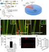
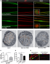
Figure 1
Figure 1. Primary screening and lace phenotype.
(A) Scheme of the screening strategy. (B) Pre-screening result with nejire RNAi with tub-GAL80ts; repo-GAL4. Survival curves were analyzed with Log-Rank Mantel Cox test. p<0.0001 (C) Results from the primary screening reveal 861 RNAi lines with lethality and 30 RNAi lines with impaired locomotion. (D) Peripheral nerves of L3 larval PNS, glial membrane (green) swelling and axonal (red) wrapping defect were observed upon knockdown of lace with 2 different RNAi lines (Transformant ID 21803, 110181). repo>mCD8-GFP/+ served as a control. Merged projection of confocal z-stacks is presented. In insets, orthogonal sections of the nerve region marked by a white line are shown (E) Quantification of the average cross-section of the peripheral nerves after glial specific knockdown of lace. (F) Merged projection of confocal z-stacks did not reveal any visible morphological changes in the neuronal morphology in the PNS after knockdown of lace specifically in the neurons (HRP). (G) Quantification of the average cross-section of the peripheral nerves after neuron-specific knockdown of lace. For the statistical analysis, unpaired t-test was performed with the respective control. Scale bar 20 µm. ns not significant. All graphs represent mean values ± SD. ** p<0.01 *** p<0.001.
Figure 2
Figure 2. lace is expressed in glial cells.
(A, B) Quantification of total glial cell number (mean±SD) upon lace knockdown in glia. Counting the number of glial nuclei in A8 peripheral nerve (n = 8) revealed no significant differences between control and lace RNAi larva. (C) Double immunolabeling of anti-β-galactosidase (red) and anti-repo (green) of lace5 (LacZ enhancer trap line) in L3 larval peripheral nerves. Colocalization shows that lace is expressed in glial cells, present in the peripheral abdominal nerves. (D) RT-PCR analysis showed that lace is expressed in the nervous system of both males and female flies. (E) Hypomorphic combination of lace mutant (lace2/lace5) showed axonal defasciculation and increase in the cross-section area of the nerve (arrows). HRP (red) and mCD8-GFP (green) were used to visualize the neuronal and glial morphology, respectively. (F) Quantification showed that the expression of UAS-lace by repo-GAL4 could rescue the mutant phenotype. Scale bar 20 µm. All graphs represent mean values ± SD. Unpaired t-test (two groups) and One-way ANOVA followed by Tukey post hoc test (for three groups) were performed for the statistical analysis. Scale 20 µm. ** p<0.01 *** p<0.001. ns not significant.
Figure 3
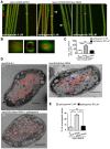
Figure 3. Wrapping glia require lace for axonal ensheathment.
(A) lace RNAi was expressed specifically in wrapping glia (Nrv2-GAL4). mCD8-GFP (green) marks the membrane of wrapping glia and HRP (red) is used to visualize the neuronal membrane. Orthogonal sections from the non-swollen region are presented as insets. Scale 20 µm. (B) TEM micrographs of L3 larval peripheral nerve cross-sections are shown. Wrapping glia is color-coded. Axonal ensheathment is incomplete upon loss of lace function in all glia (middle) or in wrapping glia (right). Proper ensheathment of axons is observed only in control (left). Scale 1 µm. (C) Quantification of the GFP signal intensity of wrapping glial membrane was performed on merged confocal projections and t-test was used for the analysis. Scale 20 µm.. (D) Quantification of the number of unwrapped axons (8–10 nerves from 4 animals for each genotype). One-way ANOVA followed by Dunnett post hoc test was performed. All graphs represent mean values ± SD. * p<0.05 *** p<0.001. (E) TUNEL assay after lace knockdown in the wrapping glia and merged projection of the peripheral nerves are presented. TUNEL (green) positive nuclei are observed only in the positive control (after DNAse addition).
Figure 4
Figure 4. Glia require CerPE for axonal ensheathment.
(A) A genetic dissection study of sphingolipid biosynthetic pathway shows that glial knockdown of Spt-I, schlank, Des1 and Pect result in swelling and wrapping defects (repo-GAL4) similar to the lace phenotype. Merged projections of confocal stacks are presented. Scale 20 µm. (B) Sphingolipid biosynthesis pathway. (C) RNAi against these 4 genes were expressed in neurons by using elav-GAL4 driver line. HRP (red) is used to visualize the membrane morphology of the neurons. Scale 20 µm. (D) Quantification (mean± SD) showed that the swellings were only observed upon glial specific knockdown but not in neuron-specific knockdown.
Figure 5
Figure 5. CerPE is very essential for axonal ensheathment by wrapping glia.
(A) RNAi against Spt-I, schlank, Des1 and Pect were expressed in wrapping glia (Nrv2-GAL4). Merged projections of the peripheral nerves along with an orthogonal projection are presented. GFP (green) was used to label the wrapping glial membrane, and HRP (red) and repo (blue) were used to label the neuronal membrane and glial nuclei, respectively. (B) TEM micrographs of L3 larval peripheral nerve cross-sections are shown. Wrapping glia is color-coded. Axonal ensheathment is incomplete upon loss of lace function in wrapping glia. Proper ensheathment of axons is observed only in control (+). Scale 1 mm. (C) Quantification of GFP signal intensity of wrapping glial membrane showed a reduction of GFP signal intensity. (D) Quantification of the number of unwrapped axons. One-way ANOVA followed by Dunnett post hoc test was performed. All graphs represent mean values 6 SD. * p,0.05, ** p,0.01, *** p,0.001.
Figure 6
Figure 6. Lipidomics analysis.
Lipidomics analysis using high-resolution shotgun mass spectrometry of dissected brains and peripheral nerves derived from L3 larvae. lace, schlank, Des1 and Pect were downregulated in both neuron and glia, L3 larval brain and peripheral nerves were dissected and the amount of sphingolipid (A), sterol (B), neutral lipid (C) and phospholipids (D) were determined. One-way ANOVA with Dunnett post hoc test was used for the statistical analysis. All graphs represent mean values ± SD. * p<0.05 ** p<0.01 *** p<0.001.
Figure 7
Figure 7. Glial inhibition of lace delays afferent spike propagation.
(A) Scheme of thoracic and abdominal parts of a dissected larva, electrodes not to scale; ag abdominal ganglion mass, se1, re1 anterior suction electrode and reference electrode, se2, re2 posterior suction electrode and reference electrode. (B) Afferent unit (top) and efferent unit (bottom) recorded simultaneously in 7th right nerve of a repo>mCD8-GFP/+ control as sketched in (A). Spike templates were generated for se2 and served as trigger events for averaging both se1 and se2. Δt conduction time; bars 50 µV and 1 ms, respectively. (C) Distribution of conduction speed recorded in repo>mCD8-GFP/+ (white bars) and repo>mCD8-GFP/lace RNAi (black bars) animals from afferent (top) and efferent (bottom) units. Horizontal lines indicate medians and their 95% confidence interval of the respective distributions. Difference of medians: * p<0.05 (two-tailed), ns not significant. n number of neurons, recorded from 20 mutant and 9 control larvae.
Figure 8
Figure 8. Sphingosine rescues the glial bulging phenotype.
(A) Addition of sphingosine to diet rescues the lace phenotype. Arrowheads indicate glial swelling region. (B) Orthogonal projections showed that dietary addition of sphingosine rescues the enwrapment defect. (C) Quantification of average cross-section area after knockdown of lace in glia and upon sphingosine treatment is shown. All graphs represent mean values ± SD. One-way ANOVA followed by Tukey post hoc test was performed for statistical analysis. Scale 20 µm (D1–D3) TEM micrographs of L3 larval peripheral nerve cross-sections are shown. Wrapping glia is color-coded. Vacuoles (blue arrowhead) are present in the wrapping glia and there is loss of membrane extension resulting in lack of proper ensheathement in repo-GAL4/lace RNAi flies as compared to control repo-GAL4/+ flies. (D3) Addition of 300 µM sphingosine to the diet (Matreya, USA) rescues the ensheathment defect. (E) Quantification of the number of unwrapped axons before and after sphingosine treatment in the in lace knocked down flies. (repo-GAL4/laceRNAi). One-way ANOVA followed by Tukey post hoc test was performed. All graphs represent mean values ±SD. *** p<0.001.

Similar articles

Cited by

References

    1. Freeman MR, Doherty J (2006) Glial cell biology in Drosophila and vertebrates. Trends Neurosci 29: 82–90. - PubMed
    1. Barres BA (2008) The mystery and magic of glia: a perspective on their roles in health and disease. Neuron 60: 430–440. - PubMed
    1. Sherman DL, Brophy PJ (2005) Mechanisms of axon ensheathment and myelin growth. Nat Rev Neurosci 6: 683–690. - PubMed
    1. Mirsky R, Jessen KR (1996) Schwann cell development, differentiation and myelination. Curr Opin Neurobiol 6: 89–96. - PubMed
    1. Kretzschmar D, Hasan G, Sharma S, Heisenberg M, Benzer S (1997) The swiss cheese mutant causes glial hyperwrapping and brain degeneration in Drosophila. J Neurosci 17: 7425–7432. - PMC - PubMed

Publication types

Substances

LinkOut - more resources