. 2013;9(12):e1004031.
doi: 10.1371/journal.pgen.1004031. Epub 2013 Dec 12.

The NuRD chromatin-remodeling enzyme CHD4 promotes embryonic vascular integrity by transcriptionally regulating extracellular matrix proteolysis

Affiliations

The NuRD chromatin-remodeling enzyme CHD4 promotes embryonic vascular integrity by transcriptionally regulating extracellular matrix proteolysis

Kyle G Ingram et al. PLoS Genet. 2013.

Abstract

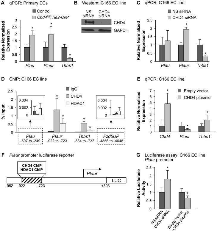
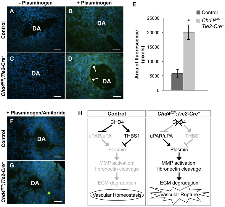
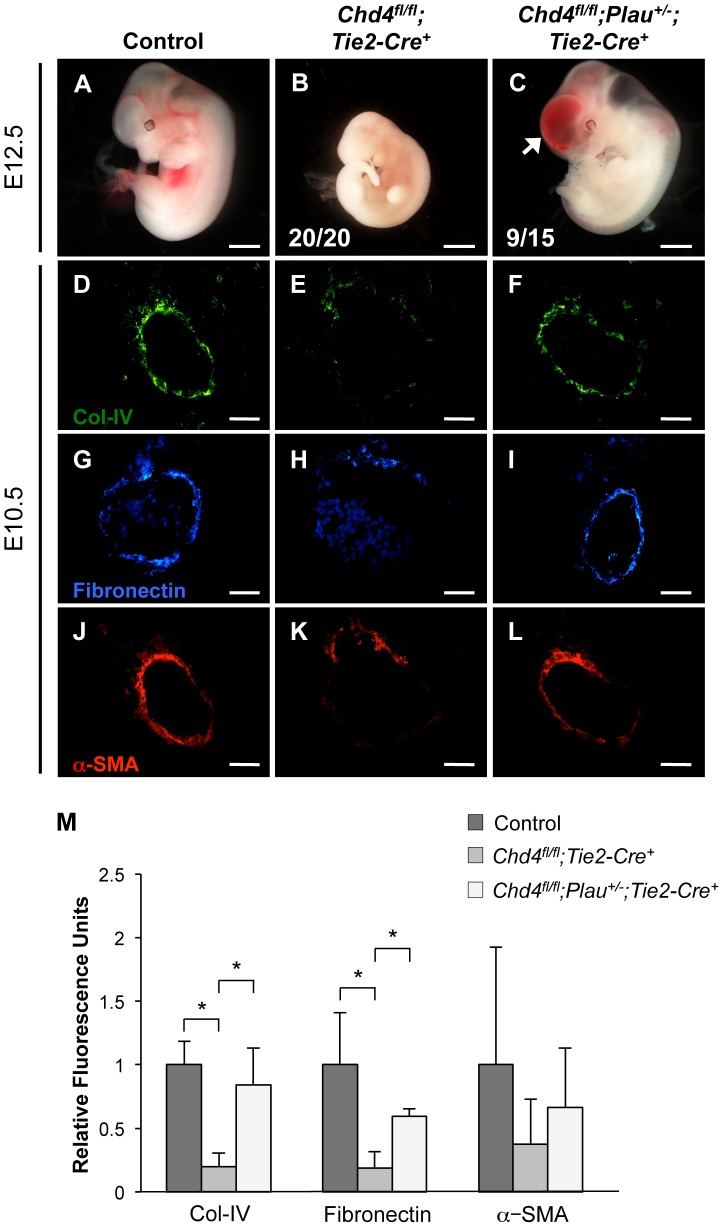
The extracellular matrix (ECM) supports vascular integrity during embryonic development. Proteolytic degradation of ECM components is required for angiogenesis, but excessive ECM proteolysis causes blood vessel fragility and hemorrhage. Little is understood about how ECM proteolysis is transcriptionally regulated during embryonic vascular development. We now show that the NuRD ATP-dependent chromatin-remodeling complex promotes vascular integrity by preventing excessive ECM proteolysis in vivo. Mice lacking endothelial CHD4--a catalytic subunit of NuRD complexes--died at midgestation from vascular rupture. ECM components surrounding rupture-prone vessels in Chd4 mutants were significantly downregulated prior to embryonic lethality. Using qPCR arrays, we found two critical mediators of ECM stability misregulated in mutant endothelial cells: the urokinase-type plasminogen activator receptor (uPAR or Plaur) was upregulated, and thrombospondin-1 (Thbs1) was downregulated. Chromatin immunoprecipitation assays showed that CHD4-containing NuRD complexes directly bound the promoters of these genes in endothelial cells. uPAR and THBS1 respectively promote and inhibit activation of the potent ECM protease plasmin, and we detected increased plasmin activity around rupture-prone vessels in Chd4 mutants. We rescued ECM components and vascular rupture in Chd4 mutants by genetically reducing urokinase (uPA or Plau), which cooperates with uPAR to activate plasmin. Our findings provide a novel mechanism by which a chromatin-remodeling enzyme regulates ECM stability to maintain vascular integrity during embryonic development.


Conflict of interest statement

The authors have declared that no competing interests exist.

Figures

Figure 1
Figure 1. Chd4fl/fl;Tie2-Cre+ embryos undergo vascular rupture by E11.25.
(A) Chd4fl/+ females were mated with Chd4fl/+;Tie2-Cre+ males, and live progeny from 22 litters were genotyped and scored at weaning. No live Chd4fl/fl;Tie2-Cre+ mice were recovered [χ2(5dof): p<0.001]. (B–E) Gross images of E11.5 littermate control (B,D) and Chd4fl/fl;Tie2-Cre+ (C,E) embryos. Arrow in panel E indicates massive hemorrhage within the ventral trunk region of a Chd4fl/fl;Tie2-Cre+ embryo. (F) Chd4fl/fl females were mated with Chd4fl/+;Tie2-Cre+ males, and dissections were performed on E10.5–12.5 embryos. Dead embryos were characterized by absence of heartbeat and onset of necrosis. No surviving Chd4fl/fl;Tie2-Cre+ embryos were found at E12.5 [χ2(3dof): p<0.01]. (G–L) Hematoxylin and eosin (H&E) staining of E10.5 (G,H) or E11.2±5 (I–L) littermate control (G,I,K) and Chd4fl/fl;Tie2-Cre+ (H,J,L) embryos. Boxed regions in panels I and J are shown at higher magnification in panels K and L respectively. Arrow in panel L reveals site of vascular rupture between the mutant dorsal aorta and cardinal vein; arrowhead indicates blood in extravascular tissues. DA, dorsal aorta; CV, cardinal vein. (M–P) Transmission electron micrographs from vessel walls of E10.5 littermate control (M,O) and Chd4fl/fl;Tie2-Cre+ (N,P) embryos. Arrowheads in panels M and N indicate smooth muscle cells adjacent to endothelial cells (ECs). Arrow in panel N points to a long, thin EC extension. (O,P) Endothelial cell junctions (*) are intact in both control and mutant sections. Scale bars: 1 mm (B–E); 100 µm (G–L); 2 µm (M,N); 500 nm (O,P).
Figure 2
Figure 2. Extracellular matrix (ECM) components are diminished around Chd4fl/fl;Tie2-Cre+ vessels prior to rupture.
Histological sections of dorsal aortae (DA) from E10.5 littermate control (A,C,E) and Chd4fl/fl;Tie2-Cre+ (B,D,F) embryos were stained with antibodies against the ECM basement membrane component type IV collagen (Col-IV; A,B) or the ECM component fibronectin (C,D) or against the smooth muscle cell differentiation marker α-SMA (E,F). Representative images from three independent experiments on separate sets of embryos are shown. Scale bars: 50 µm (A–F).
Figure 3
Figure 3. CHD4 differentially regulates Plaur and Thbs1 expression in endothelial cells.
(A) qPCR with gene-specific primers for Plau, Plaur, and Thbs1 was performed on endothelial cells isolated from E10.5 littermate control and Chd4fl/fl;Tie2-Cre+ embryos. Data were normalized to the relative expression of control samples. Error bars represent SD of results from three independent experiments. (B and C) C166 endothelial cells were transfected with nonspecific (NS) or CHD4-specific siRNA oligonucleotides for 24 h. (B) Western blot analysis was performed on cell lysates using antibodies that recognize CHD4 or GAPDH. A representative blot from 3 independent experiments is shown. (C) RNA was isolated, cDNA was synthesized, and qPCR was performed using Plau-, Plaur-, or Thbs1-specific primers. Data were normalized to the relative expression of NS siRNA-treated samples. Error bars represent SD of results from three to four independent experiments. (D) Chromatin immunoprecipitation (ChIP) assays were carried out in C166 endothelial cells using antibodies against normal mouse IgG (negative control), CHD4, or HDAC1. Immunoprecipitated DNA was analyzed by qPCR to examine CHD4 and HDAC1 enrichment at the Plau, Plaur, and Thbs1 promoters. A transcriptionally inactive region approximately 5 kb upstream of the Fzd5 transcription start site (Fzd5UP) was assessed as a negative control for CHD4 and HDAC1 binding. Data are represented as a percent of total input chromatin. Error bars represent SD of results from three independent experiments. Results for Plau and Fzd5UP ChIP experiments are magnified in the insets. For the Plaur and Thbs1 ChIPs, CHD4 and HDAC1 binding were statistically compared against IgG binding at the respective loci or against CHD4 and HDAC1 binding at the Fzd5UP locus; both sets of comparisons revealed significant enrichment. (E) qPCR with gene-specific primers for Chd4, Plaur, and Thbs1 was performed on C166 endothelial cells transfected with 0.02 ng of a CHD4 expression plasmid or the analogous empty vector backbone (control). Data were normalized to the relative expression of control samples. Error bars represent SD of results from three independent experiments. (F) Schematic of the region of the murine Plaur promoter that was cloned into a luciferase (LUC) reporter plasmid for use in G. The Plaur promoter fragment encompasses the region to which CHD4 and HDAC1 were shown to bind by ChIP in D. (G) Luciferase assays were performed in C166 cells co-transfected with 250 ng of the reporter schematized in F and 10 ng of a constitutive Renilla luciferase control plasmid. Cells were also transfected with either 10 pmol of non-specific (NS) siRNA or CHD4 siRNA oligonucleotides to knock down endogenous CHD4 or with the CHD4 expression plasmid or its relevant control (empty vector) described in E. All transfections were performed for 24 h. Ratios of relative luciferase∶renilla activity were normalized to results from the control samples. Error bars represent SD of results from four independent experiments (with triplicate samples) for the siRNA-transfected samples and from five independent experiments (with triplicate samples) for the CHD4/control plasmid-transfected samples. All statistical calculations for Figure 3 were performed using a two-tailed Student's t test (*, p<0.05).
Figure 4
Figure 4. Excessive plasmin activity is detected around rupture-prone Chd4fl/fl;Tie2-Cre+ blood vessels.
(A–D) Representative images of E10.5 littermate control (A,B) and Chd4fl/fl;Tie2-Cre+ (C,D) dorsal aortae (DA) subjected to in situ zymography for detection of plasmin activity. Unfixed sections were overlaid with quenched fluorescent casein, a plasmin substrate that fluoresces upon cleavage. In the presence of exogenous plasminogen, casein cleavage (green fluorescence) was substantially higher around Chd4fl/fl;Tie2-Cre+ versus control dorsal aortae (arrows in D versus B). The lack of significant casein cleavage in the absence of exogenous plasminogen (A and C) indicates that the casein cleavage seen in D resulted from plasmin activity. Hoechst (blue) was used for counterstaining. (E) Quantification of three independent in situ zymography experiments such as those shown in B and D from three sets of control and Chd4fl/fl;Tie2-Cre+ embryos. Fluorescence generated by casein cleavage was quantified and measured in pixels, comparing comparably sized control and Chd4fl/fl;Tie2-Cre+ dorsal aortae. Data are presented as mean ± SD. (F–G) In situ zymography for plasmin activity was performed as described in A for control and Chd4fl/fl;Tie2-Cre+ embryonic sections in the presence of the uPA inhibitor amiloride. Amiloride decreased the level of plasmin activity surrounding the Chd4fl/fl;Tie2-Cre+ DA (G) to that seen surrounding the control DA (F). (H) Model for how CHD4 impacts ECM proteolysis and vascular integrity through transcriptional regulation of Thbs1 and Plaur. (Left panel) In control endothelial cells, plasmin production and ECM degradation are curbed by CHD4-mediated inhibition of the genes encoding uPA/uPAR and activation of the gene encoding THBS1, resulting in vascular homeostasis. (Right panel) In Chd4fl/fl;Tie2-Cre+ endothelial cells, loss of CHD4 leads to increased plasmin activation, which enhances MMP activation and fibronectin cleavage. The net result is excessive ECM degradation and vascular rupture. Scale bars: 100 µm.
Figure 5
Figure 5. Genetic reduction of urokinase restores ECM components and partially rescues vascular rupture in Chd4fl/fl;Tie2-Cre+ embryos.
(A–C) Representative images of littermate control (A), Chd4fl/fl;Tie2-Cre+ (B), and Chd4fl/fl;Plau+/−;Tie2-Cre+ (C) embryos at E12.5. All Chd4fl/fl;Tie2-Cre+ embryos examined (20/20) were pale and necrotic (B). 60% of Chd4fl/fl;Plau+/−;Tie2-Cre+ embryos (9 out of 15) were comparable in size to littermate control embryos, although they displayed blood in the brain (arrow) and/or spinal cord (C). (D–L) Histological sections of dorsal aortae from E10.5 control, Chd4fl/fl;Tie2-Cre+, and Chd4fl/fl;Plau+/−;Tie2-Cre+ embryos were stained with antibodies against type IV collagen (Col-IV; D–F), fibronectin (G–I), or α-SMA (J–L). Scale bars: 1 mm (A–C); 50 µm (D–L). (M) Quantification of immunostained ECM components, such as those shown in panels D–L. Relative fluorescent intensity was measured and normalized to fluorescence in the control sections. Error bars represent SD of results from three independent experiments using three different sets of littermate embryos. Significant differences were calculated using a two-tailed Student's t test (*, p<0.05).

Similar articles

Cited by

References

    1. Davis GE, Senger DR (2005) Endothelial extracellular matrix: biosynthesis, remodeling, and functions during vascular morphogenesis and neovessel stabilization. Circ Res 97: 1093–1107. - PubMed
    1. Murakami M, Simons M (2009) Regulation of vascular integrity. J Mol Med 87: 571–582. - PMC - PubMed
    1. Sakalihasan N, Limet R, Defawe OD (2005) Abdominal aortic aneurysm. Lancet 365: 1577–1589. - PubMed
    1. Ruigrok YM, Rinkel GJ, Wijmenga C (2005) Genetics of intracranial aneurysms. Lancet Neurol 4: 179–189. - PubMed
    1. Poschl E, Schlotzer-Schrehardt U, Brachvogel B, Saito K, Ninomiya Y, et al. (2004) Collagen IV is essential for basement membrane stability but dispensable for initiation of its assembly during early development. Development 131: 1619–1628. - PubMed

Publication types

MeSH terms

Substances

LinkOut - more resources