. 2013;9(12):e1004032.
doi: 10.1371/journal.pgen.1004032. Epub 2013 Dec 12.

A domesticated PiggyBac transposase interacts with heterochromatin and catalyzes reproducible DNA elimination in Tetrahymena

Affiliations

A domesticated PiggyBac transposase interacts with heterochromatin and catalyzes reproducible DNA elimination in Tetrahymena

Alexander Vogt et al. PLoS Genet. 2013.

Abstract

The somatic genome of the ciliated protist Tetrahymena undergoes DNA elimination of defined sequences called internal eliminated sequences (IESs), which account for ~30% of the germline genome. During DNA elimination, IES regions are heterochromatinized and assembled into heterochromatin bodies in the developing somatic nucleus. The domesticated piggyBac transposase Tpb2p is essential for the formation of heterochromatin bodies and DNA elimination. In this study, we demonstrate that the activities of Tpb2p involved in forming heterochromatin bodies and executing DNA elimination are genetically separable. The cysteine-rich domain of Tpb2p, which interacts with the heterochromatin-specific histone modifications, is necessary for both heterochromatin body formation and DNA elimination, whereas the endonuclease activity of Tpb2p is only necessary for DNA elimination. Furthermore, we demonstrate that the endonuclease activity of Tpb2p in vitro and the endonuclease activity that executes DNA elimination in vivo have similar substrate sequence preferences. These results strongly indicate that Tpb2p is the endonuclease that directly catalyzes the excision of IESs and that the boundaries of IESs are at least partially determined by the combination of Tpb2p-heterochromatin interaction and relaxed sequence preference of the endonuclease activity of Tpb2p.


Conflict of interest statement

The authors have declared that no competing interests exist.

Figures
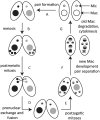
Figure 1
Figure 1. Conjugation of Tetrahymena.
Under starvation conditions, two Tetrahymena cells form a pair (A) and initiate sexual reproduction (conjugation). The Mic undergoes meiosis, and three of the four meiotic products are degraded (B), whereas the remaining product divides mitotically to form two pronuclei in each cell (C). One of the two pronuclei is exchanged with the mating partner, and fusion leads to formation of the zygotic nucleus (D). The zygotic nucleus divides mitotically twice (E); two nuclei become Mics, and the other two differentiate into Macs (F). The parental Mac is degraded. After cell division, the nuclei are distributed to the daughter cells (G).
Figure 2
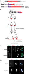
Figure 2. Construction and analyses of TPB2 conditional KO (cKO) cells.
(A) The TPB2 conditional KO construct (MTT1-TPB2) was introduced into the endogenous TPB2 locus (WT TPB2 locus) by homologous recombination to create the TPB2 conditional KO locus, in which TPB2 expression is under the control of the cadmium-inducible MTT1 promoter. (B) Germline transformation with the MTT1-TPB2 construct resulted in one heterozygous TPB2 cKO clone (F1), which was then crossed with WT to obtain the heterozygous TPB2 cKO clones with different mating types (F2). The heterozygous TPB2 cKO clones were phenotypically assorted (dotted arrows) until they lost Mac copies of MTT1-TPB2 and afterwards crossed with each other to obtain homozygous TPB2 cKO strains (F3). (C) Mating TPB2 cKO cells were incubated with (induced) or without (non-induced) cadmium, and exconjugants (progeny) were fixed at 14 hr post-mixing for immunofluorescence staining. The cells were triple stained with a guinea pig anti-Pdd1p antibody (green), which marks heterochromatin, a rabbit anti-Tpb2p antibody (red) and DAPI (blue), which stains DNA. The number of cells displaying the phenotype represented by the pictures in each culture condition are shown. (D) Mating TPB2 cKO cells were incubated with (induced) or without (non-induced) cadmium, and exconjugants (progeny) were fixed at 36 hr post-mixing for DNA FISH against the Tlr1 IES to assess DNA elimination. DNA was counterstained with DAPI. The number of cells displaying the phenotype represented by the pictures in each culture condition are shown. i = micronucleus; na = new macronucleus.
Figure 3
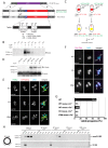
Figure 3. Functional analyses of the endonuclease and cysteine-rich domains of TPB2 in vivo.
(A) The TPB2 rescue construct (MTT2-TPB2) was introduced into the endogenous MTT1 locus (WT MTT1 locus) by homologous recombination to create the TPB2 rescue locus, in which TPB2 expression is under the control of the copper-inducible MTT2 promoter. Three different TPB2 rescue constructs encoding for wild-type Tpb2p (WT), catalytic-dead Tpb2p (CD) and a cysteine-rich mutant Tpb2p (CRM) were used. (B) TPB2 cKO strains were mated and either left uninduced or cadmium or copper were added during conjugation. Tpb2p expression from the MTT1 promoter at the TPB2 cKO locus at 9, 12 and 15 h post-mixing was detected by western blot using an anti-Tpb2p antiserum. (C) Scheme of production of the TPB2 rescue strains. A TPB2 rescue construct was introduced into the MTT1 locus of the Mac of TPB2 cKO cells. (D) TPB2 rescue strains expressing WT, CD or CRM TPB2 were mated, and Tpb2p expression from the MTT2 promoter at the rescue locus was compared between in the absence (−) or presence (+) of CuSO4 by western blot (top). As a loading control, expression of alpha-tubulin was analyzed (bottom) (E) Mating TPB2-rescued cells with wild-type (WT), catalytic-dead (CD) or cysteine-rich mutant (CRM) construct were incubated with copper, and exconjugants (progeny) were fixed at 14 hr post-mixing for immunofluorescence staining. The cells were triple stained with a guinea pig anti-Pdd1p antibody (green), which marks heterochromatin, a rabbit anti-Tpb2p antibody (red), and DAPI (blue), which stains DNA. The numbers of cells displaying the phenotype represented by the pictures in each culture condition are shown. (F) Mating TPB2 rescued cells were incubated with CuSO4, and exconjugants (progeny) were fixed at 36 hr post-mixing for DNA FISH against the Tlr1 IES to assess DNA elimination. DNA was counterstained with DAPI. The number of cells displaying the phenotype represented by the pictures in each culture condition are shown. i = micronucleus; na = new macronucleus. (G) Viability of sexual progeny of the wild-type (WT)or the TPB2 rescue strains without (−Cu2+) or with (+Cu2+) induction of the wild-type (WT rescue), catalytic-dead (CD rescue) or cysteine-rich mutant (CRM rescue) TPB2 were analyzed. 192 single mating pairs were placed into drops of medium and incubated for ∼60 h at 30°C. Completion of conjugation was confirmed by testing for negative expression of the marker specific for the parental Macs. (H) Genomic DNA was extracted from starved or mating (10, 12, 14 and 16 h post-mixing) cells of the wild-type or the TPB2 rescue strains without (−Cu2+) or with (+Cu2+) induction of the wild-type (WT), catalytic-dead (CD) or cysteine-rich mutant (CRM) rescue constructs, and circularized mse2.9- and R-IES elements were detected by nested PCR as shown on the left.
Figure 4
Figure 4. In vitro histone peptide-binding assay.
Indicated biotin-tagged histone peptides were immobilized on streptavidin Dynabeads and incubated with the recombinant expressed MBP-tagged cysteine-rich domain (566 aa–657 aa) of Tpb2p (MBP-Tpb2p-CRD (WT)) or peptides having mutations (C618A, C629A) at the putative metal-binding cysteines (MBP-Tpb2p-CRD (mut)). Proteins co-precipitated with the histone peptides were detected by western blot using anti-MBP antiserum. The average intensities of bands from three independent experiments were normalized to the value of “scrambled” peptides. Standard deviations are shown after the average intensity values. P-values from a student's T-test are shown.
Figure 5
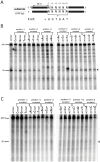
Figure 5. In vitro Tpb2p endonuclease assay.
(A) Schematic drawing of the oligo DNA substrates. The substrates were 100 bp long, and 6 bp boundary sequences from the R-IES (5′-AGTGAT-3′) or those having mutated versions of the boundary, in which every position was substituted to every other possible nucleotide, were placed at the 50th to 55th position, which were designated positions −1 to +5. The top strands of the substrates were 32P-labeled at their 5′-ends. The expected cleavage site, where a 4-base 5′-overhang DSB is expected to occur during DNA elimination in vivo, is after the 50th nucleotide. (B, C) The substrates were incubated with the wild-type Tpb2p (B) or the catalytic dead Tpb2p (Tpb2p-CD) (C) expressed recombinantly in E. coli. The products were separated in a denaturing polyacrylamide gel and visualized by autoradiography. The positions of the 50 and 100 nt markers separated in the same gel are shown on the left.
Figure 6
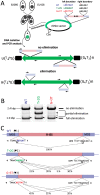
Figure 6. In vivo elimination assay using mutated R-IES boundaries.
(A) Two wild-type strains (B2086 and CU428) were mated, and the rDNA vector containing the R-IES and its flanking regions was introduced into the new Mac of their progeny. The left boundary of the R-IES was WT, the position +2 T was mutated to G (T+2G) or the position +3 G was mutated to T (G+3T). The introduced circular rDNA vector was rearranged into an rDNA “minichromosome” in which two copies of rDNA are joined in inverted orientations and telomeres are formed at the ends. The R-IES inserted in the 3′ non-coding region of the rDNA is subjected to DNA elimination similar to the endogenous R-IES . The reported cleavage positions at the endogenous R-IES locus are indicated by red arrows. (B) Twenty-four progeny from each construct were pooled, and their genomic DNA was analyzed by PCR using the primer set shown in (A) to observe the elimination of R-IES from the rDNA. The PCR products were separated by agarose gel electrophoresis. The quickly migrating products (∼1.3 kbp, marked with *1, *2 or *3) correspond with the rDNA locus where the full (or nearly full) length of R-IES was eliminated (full elimination). The most slowly migrating product (∼2.4 kbp) bands correspond with the rDNA locus where no R-IES elimination occurred (no elimination). Some products (open and closed arrowheads) migrating between the two products correspond with the rDNA locus where the R-IES were partially eliminated (partial elimination) (C) The ∼1.3-kbp PCR products, marked with *1, *2 or *3 in (B), were cloned, and sequences of 48 clones from each rDNA construct were analyzed to assign cleavage sites. The lines under the boundary sequences indicate the positions where the cleavage sites were assigned at exact positions. The brackets indicate the positions where the cleavage site could not be mapped at exact positions due to sequence redundancies (direct repeats). The fractions of the cleavage sites among the 48 observed clones are shown. The reported cleavage positions at the endogenous R-IES locus are indicated by red arrows.

Similar articles

Cited by

References

    1. Orgel LE, Crick FH (1980) Selfish DNA: the ultimate parasite. Nature 284: 604–607. - PubMed
    1. Almeida R, Allshire RC (2005) RNA silencing and genome regulation. Trends Cell Biol 15: 251–258 doi:10.1016/j.tcb.2005.03.006 - DOI - PubMed
    1. Volff J-N (2006) Turning junk into gold: domestication of transposable elements and the creation of new genes in eukaryotes. Bioessays 28: 913–922 doi:10.1002/bies.20452 - DOI - PubMed
    1. Cheng C-Y, Vogt A, Mochizuki K, Yao M-C (2010) A domesticated piggyBac transposase plays key roles in heterochromatin dynamics and DNA cleavage during programmed DNA deletion in Tetrahymena thermophila. Mol Biol Cell 21: 1753–1762 doi:10.1091/mbc.E09-12-1079 - DOI - PMC - PubMed
    1. Prescott DM (1994) The DNA of ciliated protozoa. Microbiol Rev 58: 233–267. - PMC - PubMed

Publication types

LinkOut - more resources