Review
. 2013 Dec 2:7:86.
doi: 10.3389/fnint.2013.00086.

Stress modulates intestinal secretory immunoglobulin A

Affiliations
Review

Stress modulates intestinal secretory immunoglobulin A

Rafael Campos-Rodríguez et al. Front Integr Neurosci. .

Abstract

Stress is a response of the central nervous system to environmental stimuli perceived as a threat to homeostasis. The stress response triggers the generation of neurotransmitters and hormones from the hypothalamus pituitary adrenal axis, sympathetic axis and brain gut axis, and in this way modulates the intestinal immune system. The effects of psychological stress on intestinal immunity have been investigated mostly with the restraint/immobilization rodent model, resulting in an up or down modulation of SIgA levels depending on the intensity and time of exposure to stress. SIgA is a protein complex formed by dimeric (dIgA) or polymeric IgA (pIgA) and the secretory component (SC), a peptide derived from the polymeric immunoglobulin receptor (pIgR). The latter receptor is a transmembrane protein expressed on the basolateral side of gut epithelial cells, where it uptakes dIgA or pIgA released by plasma cells in the lamina propria. As a result, the IgA-pIgR complex is formed and transported by vesicles to the apical side of epithelial cells. pIgR is then cleaved to release SIgA into the luminal secretions of gut. Down modulation of SIgA associated with stress can have negative repercussions on intestinal function and integrity. This can take the form of increased adhesion of pathogenic agents to the intestinal epithelium and/or an altered balance of inflammation leading to greater intestinal permeability. Most studies on the molecular and biochemical mechanisms involved in the stress response have focused on systemic immunity. The present review analyzes the impact of stress (mostly by restraint/immobilization, but also with mention of other models) on the generation of SIgA, pIgR and other humoral and cellular components involved in the intestinal immune response. Insights into these mechanisms could lead to better therapies for protecting against pathogenic agents and avoiding epithelial tissue damage by modulating intestinal inflammation.

Keywords: SIgA; brain-gut axis; glucocorticoids; intestinal mucosa; pIgR; restraint-stress.


Figures

FIGURE 1
FIGURE 1
In response to stress, the hypothalamus (H) releases the corticotrophin releasing factor (CRF) into the anterior pituitary (P), causing the release of adrenocorticotropic hormone (ACTH) into the blood flow. ACTH stimulates the generation of glucocorticoids (cortisol in humans and corticosterone in mice) in the cortex of the adrenal gland (A), which are then released into the blood. Stress also activates the autonomic sympathetic nerves in the medulla of the adrenal gland to elicit the production of catecholamines, norepinephrine and epinephrine, which are then released into the blood. Glucocorticoids and catecholamines influence the generation of interleukins, which are involved in the viability and proliferation of immunocompetent gut cells via receptors.
FIGURE 2
FIGURE 2
Stress also triggers the activation of the enteric nervous system, including afferent and efferent intrinsic intestinal nerves (afferent nerves send signals from periphery to the brain; efferent nerves from the brain to the periphery) and extrinsic innervations, whether sympathetic (splanchnic) or parasympathetic (Vagus nerve). The enteric nervous system is connected to the CNS via sympathetic and parasympathetic pathways, forming the brain-gut axis (BGA). Four levels for the control of BGA are shown (Wood et al., 1999). The stress response of the BGA influences the generation of dIgA and/or the pIgR mediated trancytosis. NTs, neurotransmitters; NPs, neuropeptides; GCs, glucocorticoids.
FIGURE 3
FIGURE 3
Simplified model of IgA generation. Antigen presenting cells and dendritic cells are located in the subepithelial dome (SED) beneath M cells. After sampling, luminal antigen is processed and expressed on the surface as an antigen-derived peptide associated with the MHC-II molecule. Dendritic cells with the MCH II peptide and the CD40 antigen on the surface migrate to the perifollicular zone of Peyer’s patches to interact with Th2 lymphocytes endowed with TCRs and CD40 ligand (CD40L) on their surface. Immunological synapsis between APC and Th2 cells via the binding of the MHC-II peptide to TCR and of CD40 to CD40L leads to the activation of Th2 and the release of TGF-β. Along with signals elicited by the CD40/CD40L binding, TGF-β is a Th2-derived IL essential for the class switch recombination of IgM+ B cells to generate IgA+ B cells, occurring in the germinal center of Peyer’s patches (highlighted in blue). Other Th2 ILs (e.g., IL-4, -5, -6, and -10) favor the differentiation and maturation of IgA+ B cells into IgA+ plasma cells. IgA+ B lymphocytes acquire the expression of gut-homing receptors, such as α4β7, CCR9 and CCR10, and migrate toward efferent lymphatic vessels and mesenteric lymphoid nodules. From there they can reach the blood stream and migrate to the lamina propria (the gut effector site). Gut epithelial cells express the MadCAM1 ligand for α4β7, and release CCL25 and CCL28, chemokines that are ligands for the CCR9 and CCR10 receptors, respectively.
FIGURE 4
FIGURE 4
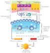
In the gut lamina propria, IgA+ B cells further differentiate into IgA+ plasma cells that secrete dIgA or pIgA antibodies joined by the J chain. dIgA or pIgA are captured by the polymeric immunoglobulin receptor (pIgR), a transmembrane receptor with five extracellular domains and an intracellular tail expressed at the basolateral side of the enterocytes. The dIgA-pIgR complex is internalized and transported by transcytosis to the apical side, and the extracellular portion of pIgR with five domains is proteolytically cleaved from the transmembrane region. The former is then released to the gut lumen as secretory component (SC) bound to dIgA to yield secretory IgA (SIgA). SIgA and SC secreted into the mucus layer prevent the direct adhesion to the epithelium of pathogenic agents, which are eventually cleared from the lumen. Apart from enterocytes, other cell components of the gut epithelium include enterochromaffin cells with granules of neurotransmitters, Paneth cells with granules containing defensins and lysozyme, and goblet cells with mucin granules and IEL.

References

    1. Asano M., Komiyama K. (2011). Polymeric immunoglobulin receptor. J. Oral Sci. 53 147–15610.2334/josnusd.53.147 - DOI - PubMed
    1. Bauer M. E., Perks P., Lightman S. L., Shanks N. (2001). Are adhesion molecules involved in stress-induced changes in lymphocyte distribution? Life Sci. 69 1167–117910.1016/S0024-3205(01)01200-0 - DOI - PubMed
    1. Blum J. S., Wearsch P. A., Cresswell P. (2013). Pathways of antigen processing. Annu. Rev. Immunol. 31 443–47310.1146/annurev-immunol-032712-095910 - DOI - PMC - PubMed
    1. Bodnar R. J. (2012). Endogenous opiates and behavior: 2011. Peptides 38 463–52210.1016/j.peptides.2012.09.027 - DOI - PubMed
    1. Brandtzaeg P. (2009). Mucosal immunity: induction, dissemination, and effector functions. Scand. J. Immunol. 70 505–51510.1111/j.1365-3083.2009.02319.x - DOI - PubMed