Review
. 2013 Nov 29:4:356.
doi: 10.3389/fmicb.2013.00356. eCollection 2013.

Gimme shelter: how Vibrio fischeri successfully navigates an animal's multiple environments

Affiliations
Review

Gimme shelter: how Vibrio fischeri successfully navigates an animal's multiple environments

Allison N Norsworthy et al. Front Microbiol. .

Abstract

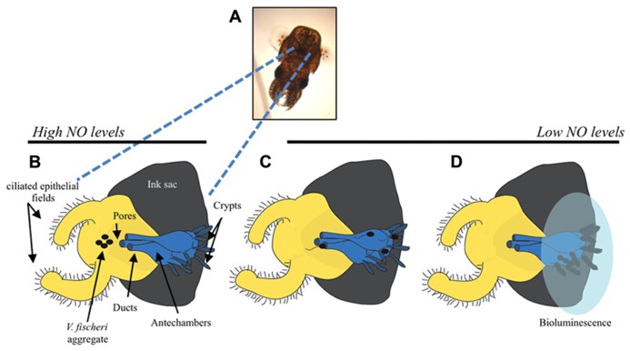
Bacteria successfully colonize distinct niches because they can sense and appropriately respond to a variety of environmental signals. Of particular interest is how a bacterium negotiates the multiple, complex environments posed during successful infection of an animal host. One tractable model system to study how a bacterium manages a host's multiple environments is the symbiotic relationship between the marine bacterium, Vibrio fischeri, and its squid host, Euprymna scolopes. V. fischeri encounters many different host surroundings ranging from initial contact with the squid to ultimate colonization of a specialized organ known as the light organ. For example, upon recognition of the squid, V. fischeri forms a biofilm aggregate outside the light organ that is required for efficient colonization. The bacteria then disperse from this biofilm to enter the organ, where they are exposed to nitric oxide, a molecule that can act as both a signal and an antimicrobial. After successfully managing this potentially hostile environment, V. fischeri cells finally establish their niche in the deep crypts of the light organ where the bacteria bioluminesce in a pheromone-dependent fashion, a phenotype that E. scolopes utilizes for anti-predation purposes. The mechanism by which V. fischeri manages these environments to outcompete all other bacterial species for colonization of E. scolopes is an important and intriguing question that will permit valuable insights into how a bacterium successfully associates with a host. This review focuses on specific molecular pathways that allow V. fischeri to establish this exquisite bacteria-host interaction.

Keywords: Euprymna scolopes; Vibrio fischeri; antimicrobials; biofilm; bioluminescence; chemotaxis; symbiosis.


Figures

FIGURE 1
FIGURE 1
Steps of E. scolopes colonization by the luminescent bacterium, V. fischeri. (A) Image of a juvenile E. scolopes. The bi-lobed light organ can be seen as a black structure in the mantle cavity. (B) Cartoon depicting one lobe of the light organ with the ink sac (gray), ciliated epithelial cells (yellow), and internal regions of the light organ (blue). Before the initial contact with V. fischeri (black ovals), E. scolopes produces the reactive nitrogen radical, nitric oxide (NO), which it subsequently down-regulates after exposure to the bacteria. Initiation of colonization requires that V. fischeri cells form a biofilm-like aggregate around the pores to the light organ. Motility is not required for biofilm formation. (C) After aggregation, V. fischeri cells utilize flagella to migrate into the pores, through the ducts and antechamber, and to establish their niche in the crypt spaces. (D) Once in the crypts, V. fischeri lose their flagella and grow to a sufficient cellular density that allows for the induction of bioluminescence genes (transparent blue oval represents luminescence). Figure modified from Nyholm and McFall-Ngai (2004).
FIGURE 2
FIGURE 2
Regulation of biofilm formation in V. fischeri. Two-component regulators control the production of the Syp biofilm. RscS and SypF are proposed to function as sensor kinases, resulting in the phosphorylation of the two downstream response regulators, SypE and SypG. SypG functions as a transcription factor to control expression of the syp locus at its four promoters, while SypE functions downstream of syp transcription to control the phosphorylation state of the small STAS domain protein, SypA. SypF is also predicted to control the activity of a RR VpsR putatively involved in cellulose biosynthesis. Biofilms can be assessed in vitro as a wrinkled colony on an agar plate, or in vivo as a bacterial aggregate that forms on the surface of the light organ. Adapted from (Visick, 2009).
FIGURE 3
FIGURE 3
Predicted flagellar synthesis pathway in V. fischeri. The V. fischeri model of flagella gene regulation is based on the pathway elucidated in the related microbe, V. cholerae (Prouty et al., 2001). Class I consists solely of the regulator, FlrA, which, together with σ54, controls expression of Class II genes. Class II proteins include both those necessary for building the base of the flagellum and also the regulators FlrB, FlrC and σ28(FliA). FlrB and FlrC control transcription of Class III genes necessary for synthesis of the distal basal body, hook, and filament, while σ28 regulates transcription of Class IV genes involved in the production of motor proteins and other miscellaneous factors. Regulators in red indicate they are important for V. fischeri to colonize the squid (Millikan and Ruby, 2003, 2004; Hussa et al., 2007; Brennan et al., 2013b).
FIGURE 4
FIGURE 4
Predicted chemotaxis pathway in V. fischeri. Methyl-accepting chemotaxis proteins (MCPs) recognize specific molecules found in the environment. A MCP is often physically linked to the sensor kinase, CheA, through the CheW protein. Ligand recognition by the MCP leads to a change in the activity of CheA. Binding of an attractant, such as (GlcNAc)2, inhibits CheA kinase activity resulting in a “run.” Conversely, interaction with a repellant promotes CheA autophosphorylation wherein the phosphoryl groups are donated to both CheB and CheY. Phospho-CheY binds to the base of the flagellar motor and causes the flagellum to switch from a counter-clockwise to clockwise rotation. This causes tumbling. The methylesterase, CheB, and the methyltransferase, CheR, both control the methylation state of the MCP allowing a cell to adapt to varying concentrations of chemicals within a chemogradient. Regulators indicated in yellow have been demonstrated to be important for squid colonization (Hussa et al., 2007; Deloney-Marino and Visick, 2012; Mandel et al., 2012).
FIGURE 5
FIGURE 5
Reactive oxygen species and reactive nitrogen species pathways and V. fischeri proteins potentially involved in modulating synthesis of these antimicrobials. ROS and RNS are indicated in red (Fang, 2004; Bowman et al., 2011). V. fischeri enzymes that have been demonstrated (solid line) or predicted (dashed line) to modulate levels of potential antimicrobial molecules are indicated in blue.
FIGURE 6
FIGURE 6
Lux pathway controlling bioluminescence in V. fischeri. At low cell density, the sensor kinases AinR and LuxP/Q are predicted to exhibit net kinase activity leading to the phosphorylation of LuxU and subsequent phosphotransfer to LuxO. Phospho-LuxO induces the expression of the inhibitory sRNA, qrr1, which leads to the degradation of litR mRNA. LitR is the transcriptional activator of luxR, which encodes a protein required for expression of the luxCDEBAG operon. Thus, at low cell density, litR translation is inhibited and the cells do not produce high levels of light. At high cell density, two distinct autoinducer molecules made by AinS (C8-HSL, diamonds) and LuxS (AI-2, circles) are predicted to be at sufficient concentrations to switch the activity of the SKs from net kinase to net phosphatase activity. This leads to dephosphorylation of the downstream regulators, litR translation, and transcription of luxR. LuxR, in conjunction with the autoinducer produced by LuxI (3-oxo-C6-HSL, triangles), leads to the transcription of the lux operon and bioluminescence (reviewed in Stabb et al., 2008). LuxR is also predicted to weakly bind to C8-HSL, which allows for the initiation of luxCDABEG expression. See text for caveats to this model.

References

    1. Altura M. A., Heath-Heckman E. A., Gillette A., Kremer N., Krachler A. M., Brennan C., et al. (2013). The first engagement of partners in the Euprymna scolopes-Vibrio fischeri symbiosis is a two-step process initiated by a few environmental symbiont cells. Environ. Microbiol. 15 2937–2950 10.1111/1462-2920.12179 - DOI - PMC - PubMed
    1. Altura M. A., Stabb E., Goldman W., Apicella M., Mcfall-Ngai M. J. (2011). Attenuation of host NO production by MAMPs potentiates development of the host in the squid-vibrio symbiosis. Cell Microbiol. 13 527–537 10.1111/j.1462-5822.2010.01552.x - DOI - PMC - PubMed
    1. Baudouin E., Pauly N., Puppo A. (2007). “Nitric oxide in nitrogen-fixing symbiosis,” in Nitric Oxide in Plant Growth, Development, and Stress Physiology, eds Lamattina L., Polacco J. (Berlin: Springer; ) 173–186
    1. Berg H. C. (2003). The rotary motor of bacterial flagella. Annu. Rev. Biochem. 72 19–54 10.1146/annurev.biochem.72.121801.161737 - DOI - PubMed
    1. Boettcher K. J., Ruby E. G. (1990). Depressed light emission by symbiotic Vibrio fischeri of the sepiolid squid Euprymna scolopes. J. Bacteriol. 172 3701–3706 - PMC - PubMed

LinkOut - more resources