

Small intestinal tumours
- PMID: 24348540
- PMCID: PMC3855980
- DOI: 10.1155/2013/702536
Small intestinal tumours
Abstract
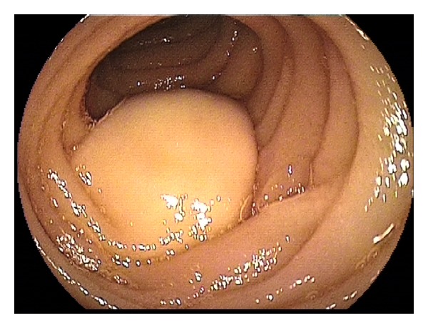
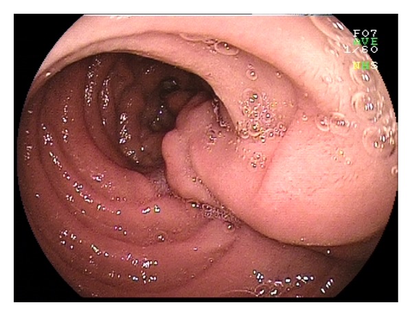
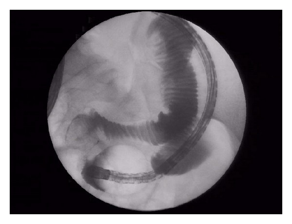
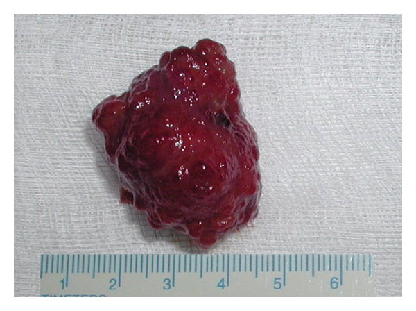
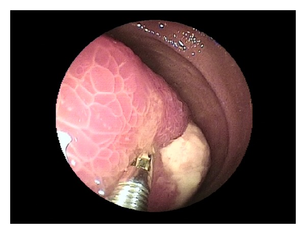
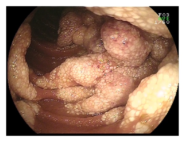
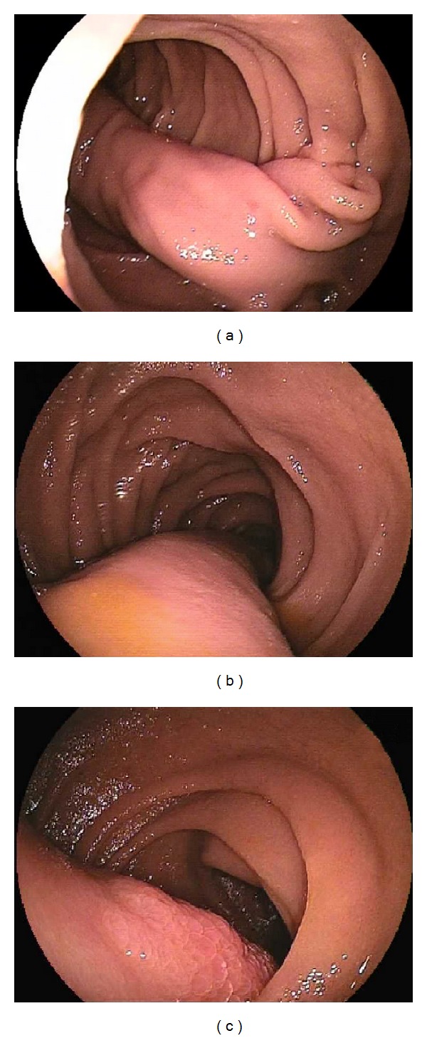
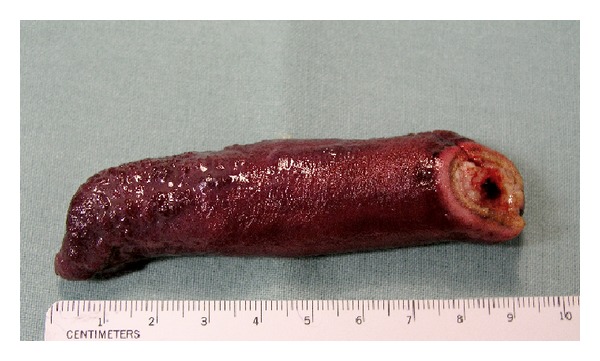
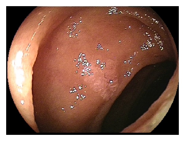
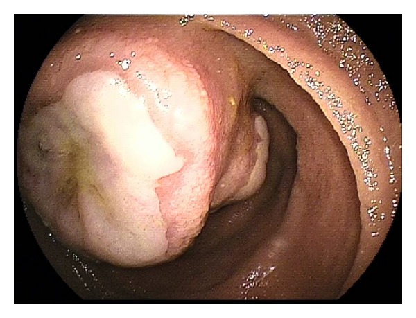
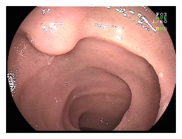
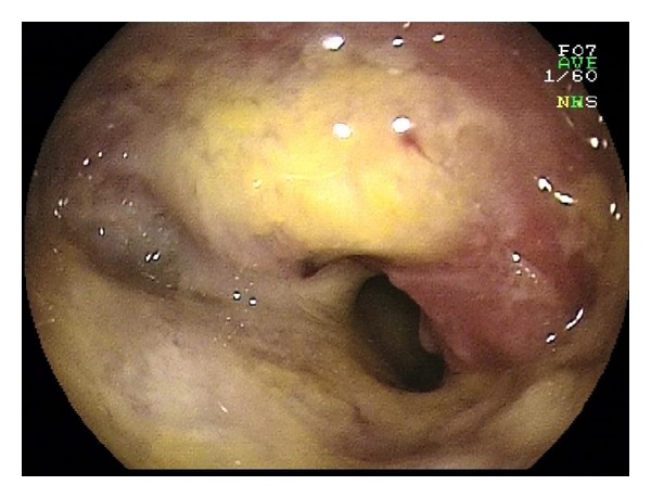
Objective. Balloon enteroscopy (BE) and capsule enteroscopy (CE) are enteroscopy methods that allow examination and treatment of the small bowel. Before the CE and BE era, the small intestine was difficult to access for investigation. Small intestinal tumours are infrequent conditions, but about half of them are malignant. Materials and Methods. A total of 303 BEs were performed in 179 patients. Oral insertion was performed in 240 and anal in 63 BEs. Indications for the procedure in our patients with small bowel tumours were anaemia and/or bleeding, obstruction, suspicion of carcinoid tumour, or suspicion of Peutz-Jeghers syndrome. Results. In 50 of our 179 patients (28%), we diagnosed some small intestinal tumours: hamartomas in Peutz-Jeghers syndrome in 16 patients, adenocarcinoma in 7, lymphoma in 6, carcinoid tumour in 4, melanoma and stromal tumour in 3, adenoma, lipoma, and inflammatory polyps in 2, and granular cell tumour, cavernous lymphangioma, fibrolipoma, Cronkhite-Canada polyps, and metastatic involvement in individual cases. Conclusion. BE facilitates exploration and treatment of the small intestine. The procedure is generally safe and useful. BE and CE are essential modalities for the management of small intestinal diseases.
Figures
Similar articles
-
Endoscopic resection of Peutz-Jeghers polyps throughout the small intestine at double-balloon enteroscopy without laparotomy.Gastrointest Endosc. 2005 Jan;61(1):140-7. doi: 10.1016/s0016-5107(04)02457-5. Gastrointest Endosc. 2005. PMID: 15672077
-
Small bowel polyp resection using device-assisted enteroscopy in Peutz-Jeghers Syndrome: Results of a specialised tertiary care centre.United European Gastroenterol J. 2020 Mar;8(2):204-210. doi: 10.1177/2050640619874525. Epub 2019 Sep 10. United European Gastroenterol J. 2020. PMID: 32213068 Free PMC article.
-
Comparison of intraoperative enteroscopy and double-balloon enteroscopy for the diagnosis and treatment of Peutz-Jeghers syndrome.Surg Endosc. 2010 Aug;24(8):1904-10. doi: 10.1007/s00464-009-0868-6. Epub 2010 Jan 28. Surg Endosc. 2010. PMID: 20108144
-
Peutz-Jeghers syndrome: diagnostic and therapeutic approach.World J Gastroenterol. 2009 Nov 21;15(43):5397-408. doi: 10.3748/wjg.15.5397. World J Gastroenterol. 2009. PMID: 19916169 Free PMC article. Review.
-
Intraoperative enteroscopy.Gastrointest Endosc Clin N Am. 2009 Jul;19(3):371-9. doi: 10.1016/j.giec.2009.04.011. Gastrointest Endosc Clin N Am. 2009. PMID: 19647646 Review.
Cited by
-
Small bowel gastrointestinal stromal tumor: a retrospective study of 32 cases at a single center and review of the literature.Ther Clin Risk Manag. 2018 Aug 22;14:1467-1481. doi: 10.2147/TCRM.S167248. eCollection 2018. Ther Clin Risk Manag. 2018. PMID: 30174429 Free PMC article.
-
De novo distal terminal ileum adenocarcinoma mimicking Crohn's disease and diagnostic challenges in imaging: a case series.BJR Case Rep. 2021 Jun 24;7(6):20210103. doi: 10.1259/bjrcr.20210103. eCollection 2022 Mar. BJR Case Rep. 2021. PMID: 35300226 Free PMC article.
-
Impact of enteroscopy on diagnosis and management of small bowel tumors.Chin J Cancer Res. 2020 Jun;32(3):319-333. doi: 10.21147/j.issn.1000-9604.2020.03.04. Chin J Cancer Res. 2020. PMID: 32694897 Free PMC article.
-
Squamous cell carcinoma of the small intestine: a case report and review of literature.Front Oncol. 2025 Apr 30;15:1550917. doi: 10.3389/fonc.2025.1550917. eCollection 2025. Front Oncol. 2025. PMID: 40371228 Free PMC article.
-
Influence of In Vitro Digestion on Composition, Bioaccessibility and Antioxidant Activity of Food Polyphenols-A Non-Systematic Review.Nutrients. 2020 May 13;12(5):1401. doi: 10.3390/nu12051401. Nutrients. 2020. PMID: 32414132 Free PMC article. Review.
References
-
- Chow WH, Linet MS, McLaughlin JK, Hsing AW, Chien HTC, Blot WJ. Risk factors for small intestine cancer. Cancer Causes and Control. 1993;4(2):163–169. - PubMed
LinkOut - more resources
Full Text Sources
Other Literature Sources
Molecular Biology Databases
