

The role of s-nitrosylation and s-glutathionylation of protein disulphide isomerase in protein misfolding and neurodegeneration
- PMID: 24348565
- PMCID: PMC3852308
- DOI: 10.1155/2013/797914
The role of s-nitrosylation and s-glutathionylation of protein disulphide isomerase in protein misfolding and neurodegeneration
Abstract
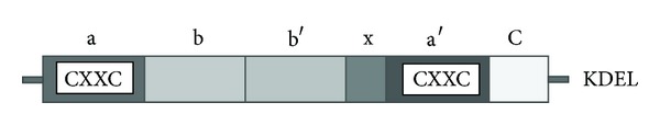
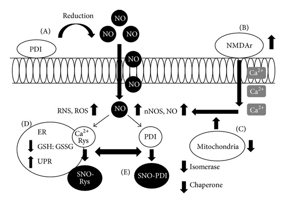
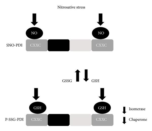
Neurodegenerative diseases involve the progressive loss of neurons, and a pathological hallmark is the presence of abnormal inclusions containing misfolded proteins. Although the precise molecular mechanisms triggering neurodegeneration remain unclear, endoplasmic reticulum (ER) stress, elevated oxidative and nitrosative stress, and protein misfolding are important features in pathogenesis. Protein disulphide isomerase (PDI) is the prototype of a family of molecular chaperones and foldases upregulated during ER stress that are increasingly implicated in neurodegenerative diseases. PDI catalyzes the rearrangement and formation of disulphide bonds, thus facilitating protein folding, and in neurodegeneration may act to ameliorate the burden of protein misfolding. However, an aberrant posttranslational modification of PDI, S-nitrosylation, inhibits its protective function in these conditions. S-nitrosylation is a redox-mediated modification that regulates protein function by covalent addition of nitric oxide- (NO-) containing groups to cysteine residues. Here, we discuss the evidence for abnormal S-nitrosylation of PDI (SNO-PDI) in neurodegeneration and how this may be linked to another aberrant modification of PDI, S-glutathionylation. Understanding the role of aberrant S-nitrosylation/S-glutathionylation of PDI in the pathogenesis of neurodegenerative diseases may provide insights into novel therapeutic interventions in the future.
Figures
References
-
- Soto C. Unfolding the role of protein misfolding in neurodegenerative diseases. Nature Reviews Neuroscience. 2003;4(1):49–60. - PubMed
-
- Glenner GG, Wong CW. Alzheimer’s disease: initial report of the purification and characterization of a novel cerebrovascular amyloid protein. 1984. Biochemical and Biophysical Research Communications. 2012;425(3):534–539. - PubMed
-
- Spillantini MG, Schmidt ML, Lee VM-Y, Trojanowski JQ, Jakes R, Goedert M. α-synuclein in Lewy bodies. Nature. 1997;388(6645):839–840. - PubMed
Publication types
LinkOut - more resources
Full Text Sources
Other Literature Sources
