

Differentiation of dental pulp stem cells into neuron-like cells in serum-free medium
- PMID: 24348580
- PMCID: PMC3852491
- DOI: 10.1155/2013/250740
Differentiation of dental pulp stem cells into neuron-like cells in serum-free medium
Abstract
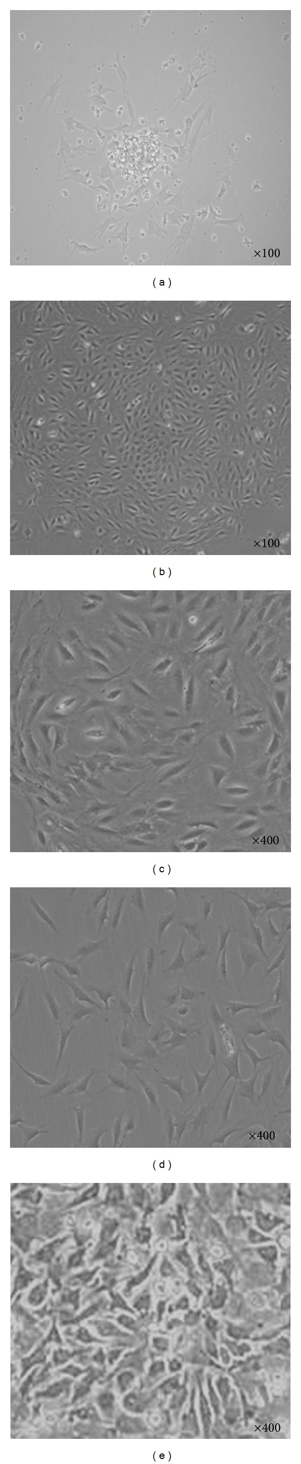
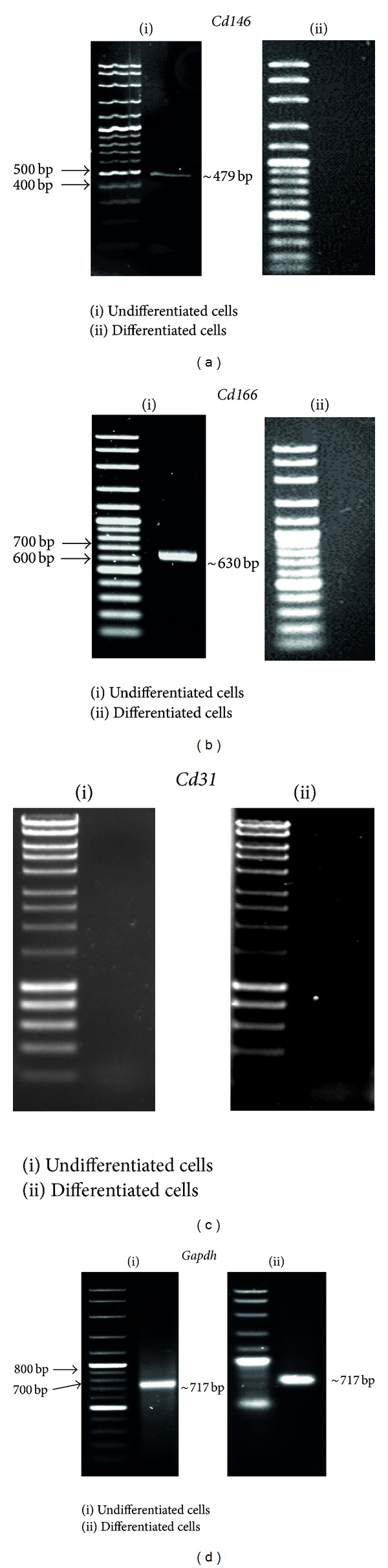
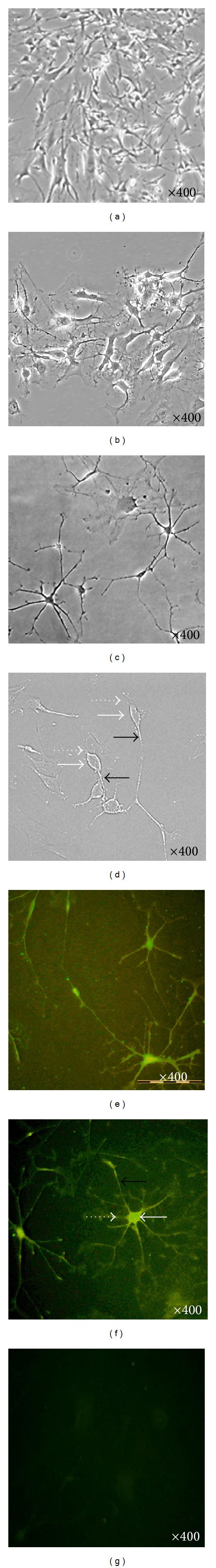
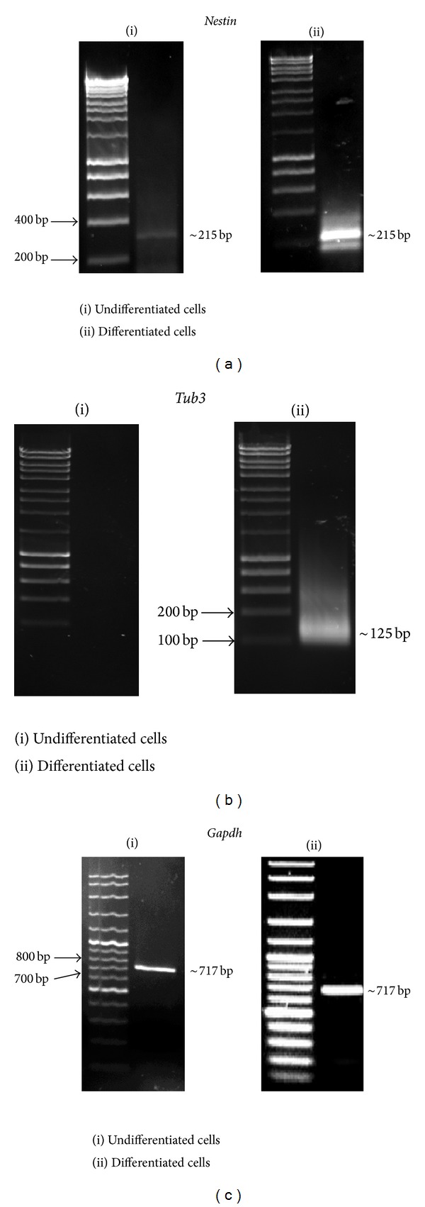
Dental pulp tissue contains dental pulp stem cells (DPSCs). Dental pulp cells (also known as dental pulp-derived mesenchymal stem cells) are capable of differentiating into multilineage cells including neuron-like cells. The aim of this study was to examine the capability of DPSCs to differentiate into neuron-like cells without using any reagents or growth factors. DPSCs were isolated from teeth extracted from 6- to 8-week-old mice and maintained in complete medium. The cells from the fourth passage were induced to differentiate by culturing in medium without serum or growth factors. RT-PCR molecular analysis showed characteristics of Cd146(+) , Cd166(+) , and Cd31(-) in DPSCs, indicating that these cells are mesenchymal stem cells rather than hematopoietic stem cells. After 5 days of neuronal differentiation, the cells showed neuron-like morphological changes and expressed MAP2 protein. The activation of Nestin was observed at low level prior to differentiation and increased after 5 days of culture in differentiation medium, whereas Tub3 was activated only after 5 days of neuronal differentiation. The proliferation of the differentiated cells decreased in comparison to that of the control cells. Dental pulp stem cells are induced to differentiate into neuron-like cells when cultured in serum- and growth factor-free medium.
Figures

Similar articles
-
Identification of neurospheres generated from human dental pulp stem cells in xeno-/serum-free conditions.Regen Ther. 2020 Feb 17;14:128-135. doi: 10.1016/j.reth.2019.11.006. eCollection 2020 Jun. Regen Ther. 2020. PMID: 32099873 Free PMC article.
-
Differentiation capacity of mouse dental pulp stem cells into osteoblasts and osteoclasts.Cell J. 2014 Feb 3;16(1):31-42. Epub 2014 Feb 3. Cell J. 2014. PMID: 24518973 Free PMC article.
-
Investigation of dental pulp stem cells isolated from discarded human teeth extracted due to aggressive periodontitis.Biomaterials. 2014 Nov;35(35):9459-72. doi: 10.1016/j.biomaterials.2014.08.003. Epub 2014 Aug 27. Biomaterials. 2014. PMID: 25172527
-
Differentiation of dental pulp stem cells into neuron-like cells.Int J Neurosci. 2020 Feb;130(2):107-116. doi: 10.1080/00207454.2019.1664518. Epub 2019 Oct 10. Int J Neurosci. 2020. PMID: 31599165
-
Characterisation of dental pulp stem cells: a new horizon for tissue regeneration?Arch Oral Biol. 2012 Nov;57(11):1439-58. doi: 10.1016/j.archoralbio.2012.08.010. Epub 2012 Sep 14. Arch Oral Biol. 2012. PMID: 22981360 Review.
Cited by
-
Hypoxia Induces DPSC Differentiation versus a Neurogenic Phenotype by the Paracrine Mechanism.Biomedicines. 2022 May 3;10(5):1056. doi: 10.3390/biomedicines10051056. Biomedicines. 2022. PMID: 35625792 Free PMC article.
-
Rapid differentiation of human dental pulp stem cells to neuron-like cells by high K+ stimulation.Biophys Physicobiol. 2020 Sep 26;17:132-139. doi: 10.2142/biophysico.BSJ-2020023. eCollection 2020. Biophys Physicobiol. 2020. PMID: 33240740 Free PMC article.
-
Exploring the neurogenic differentiation of human dental pulp stem cells.PLoS One. 2022 Nov 4;17(11):e0277134. doi: 10.1371/journal.pone.0277134. eCollection 2022. PLoS One. 2022. PMID: 36331951 Free PMC article.
-
The temporospatial relationship between mouse dental pulp stem cells and tooth innervation.J Dent Sci. 2024 Apr;19(2):1075-1082. doi: 10.1016/j.jds.2024.02.007. Epub 2024 Feb 12. J Dent Sci. 2024. PMID: 38618089 Free PMC article.
-
Analysis and comparisons of gene expression changes in patient- derived neurons from ROHHAD, CCHS, and PWS.Front Pediatr. 2023 May 10;11:1090084. doi: 10.3389/fped.2023.1090084. eCollection 2023. Front Pediatr. 2023. PMID: 37234859 Free PMC article.
References
-
- Kerkis I, Kerkis A, Dozortsev D, et al. Isolation and characterization of a population of immature dental pulp stem cells expressing OCT-4 and other embryonic stem cell markers. Cells Tissues Organs. 2007;184(3-4):105–116. - PubMed
-
- Javazon E, Tebbets J, Beggs K. Isolation, expansion and characterization of murine adult bone marrow derived mesenchymal stem cells. Blood. 2003;102:180B–181B. - PubMed
LinkOut - more resources
Full Text Sources
Other Literature Sources
Research Materials
