Case Reports
. 2013 Sep;10(3):152-5.
doi: 10.5812/iranjradiol.7108. Epub 2013 Aug 30.

A rare case of rib osteoblastoma: imaging features and review of literature

Affiliations
Case Reports

A rare case of rib osteoblastoma: imaging features and review of literature

Shokouh Taghipour Zahir et al. Iran J Radiol. 2013 Sep.

Abstract

Osteoblastoma is a rare benign, but locally aggressive bone tumor with rare malignant transformation. It mostly affects the vertebral column and long bones. Radiographically, it is seen as an expansile, oval, sclerotic or lytic mass-like lesion with well-defined borders, although sometimes it may mimic a malignant tumor such as osteogenic sarcoma by its irregular borders. Herein, we report a case of osteoblastoma in a 22 year-old man with a long history of back and neck pain accompanied with neck stiffness. On the routine chest X-ray, the salient lesion appeared as an expansile, oval, sclerotic mass with well-defined borders and speckled calcification without any internal lucency and periosteal reaction, involving the posterolateral aspect of the first left thoracic rib, a rare anatomical site. Despite the unusual location, osteoblastoma should be considered in the differential diagnosis of a solitary rib lesion.

Keywords: Bone Neoplasms; Osteoblastoma; Osteoma, Osteoid; Ribs.


Figures

Figure 1.
Figure 1.
Chest X-ray of the patient shows a well-defined salient osseous expansion with spongy-form sclerotic areas on the posterolateral side of the first left rib.
Figure 2.
Figure 2.
Non-contrast enhancement CT demonstrates an oval sclerotic, expansile lesion with spongy-form speckled calcification and a well-demarcated lesion involving the posterolateral side of the left first rib.
Figure 3.
Figure 3.
A bone trabeculae with a central nidus consisting of anastomosing bony trabeculae in a highly vascularized connective tissue (×20).
Figure 4.
Figure 4.
Variable osteoid calcifications lined by plump osteoblasts are also visible that are characteristic of osteoblastoma (×20).
Figure 5.
Figure 5.
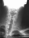
Neck and shoulder X-ray demonstrates geographic and well-circumscribed opacities.
Figure 6.
Figure 6.
MRI reveals extensive edema around the lesion and high signal intensities on T2WI with punctate low signal areas consistent with mineralization on T1WI.
Figure 7.
Figure 7.
A, Axial fast spin-echo T2W image. B, Sagittal fast spin-echo T1W image. C, Post-gadolinium coronal T1W image

References

    1. Tugcu B, Gunaldi O, Gunes M, Tanriverdi O, Bilgic B. Osteoblastoma of the temporal bone: a case report. Minim Invasive Neurosurg. 2008;51(5):310–2. doi: 10.1055/s-0028-1083816. - DOI - PubMed
    1. Mortazavi SM, Wenger D, Asadollahi S, Shariat Torbaghan S, Unni KK, Saberi S. Periosteal osteoblastoma: report of a case with a rare histopathologic presentation and review of the literature. Skeletal Radiol. 2007;36(3):259–64. doi: 10.1007/s00256-006-0169-2. - DOI - PubMed
    1. Berry M, Mankin H, Gebhardt M, Rosenberg A, Hornicek F. Osteoblastoma: a 30-year study of 99 cases. J Surg Oncol. 2008;98(3):179–83. doi: 10.1002/jso.21105. - DOI - PubMed
    1. Garcia RA, Inwards CY, Unni KK. Benign bone tumors--recent developments. Semin Diagn Pathol. 2011;28(1):73–85. - PubMed
    1. Ye J, Liu L, Wu J, Wang S. Osteoblastoma of the rib with CT and MR imaging: a case report and literature review. World J Surg Oncol. 2012;10:49. doi: 10.1186/1477-7819-10-49. - DOI - PMC - PubMed

Publication types

LinkOut - more resources